Review
. 2018 Mar;5(1):R1-R16.
doi: 10.1530/ERP-17-0062. Epub 2018 Feb 5.

EDUCATIONAL SERIES IN CONGENITAL HEART DISEASE: Echocardiographic assessment of left to right shunts: atrial septal defect, ventricular septal defect, atrioventricular septal defect, patent arterial duct

Affiliations
Review

EDUCATIONAL SERIES IN CONGENITAL HEART DISEASE: Echocardiographic assessment of left to right shunts: atrial septal defect, ventricular septal defect, atrioventricular septal defect, patent arterial duct

Antigoni Deri et al. Echo Res Pract. 2018 Mar.

Abstract

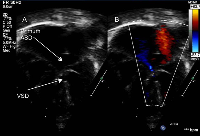
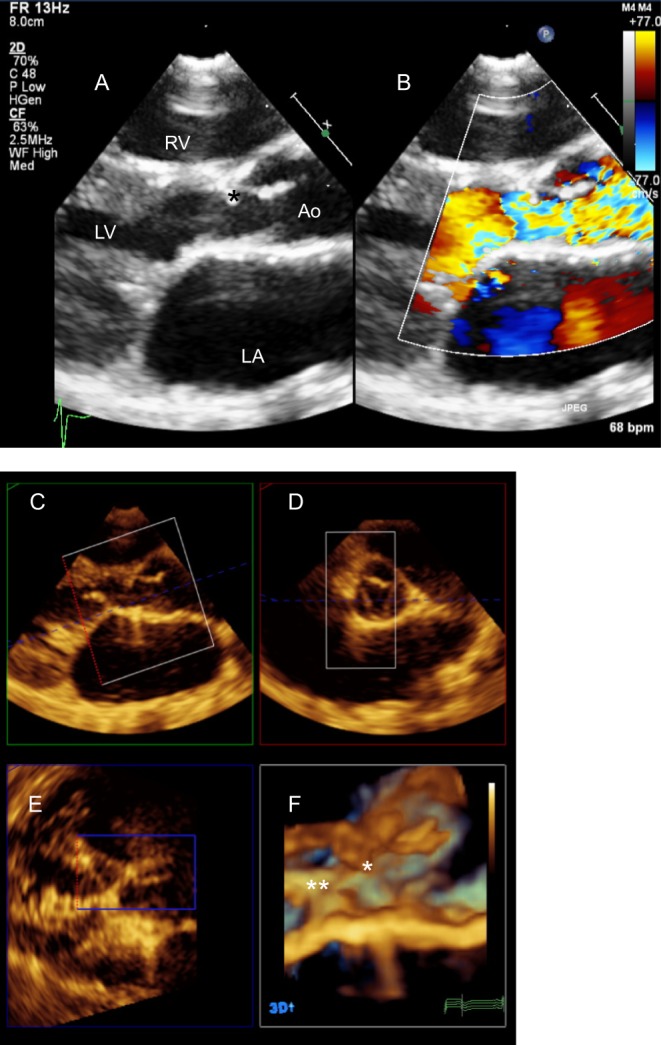
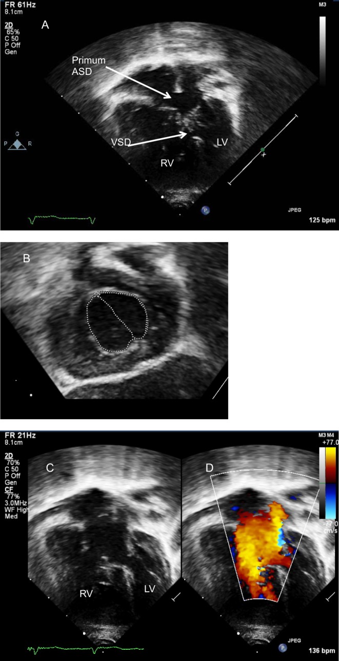
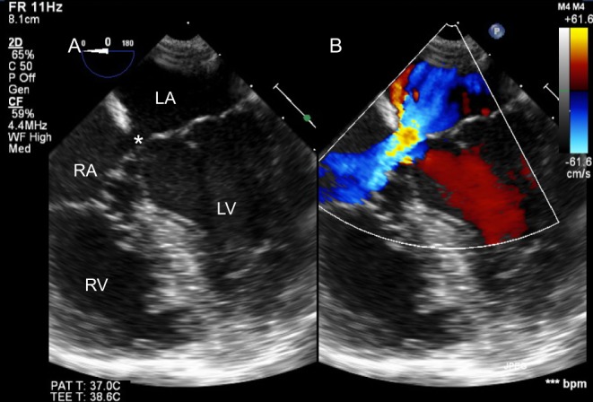
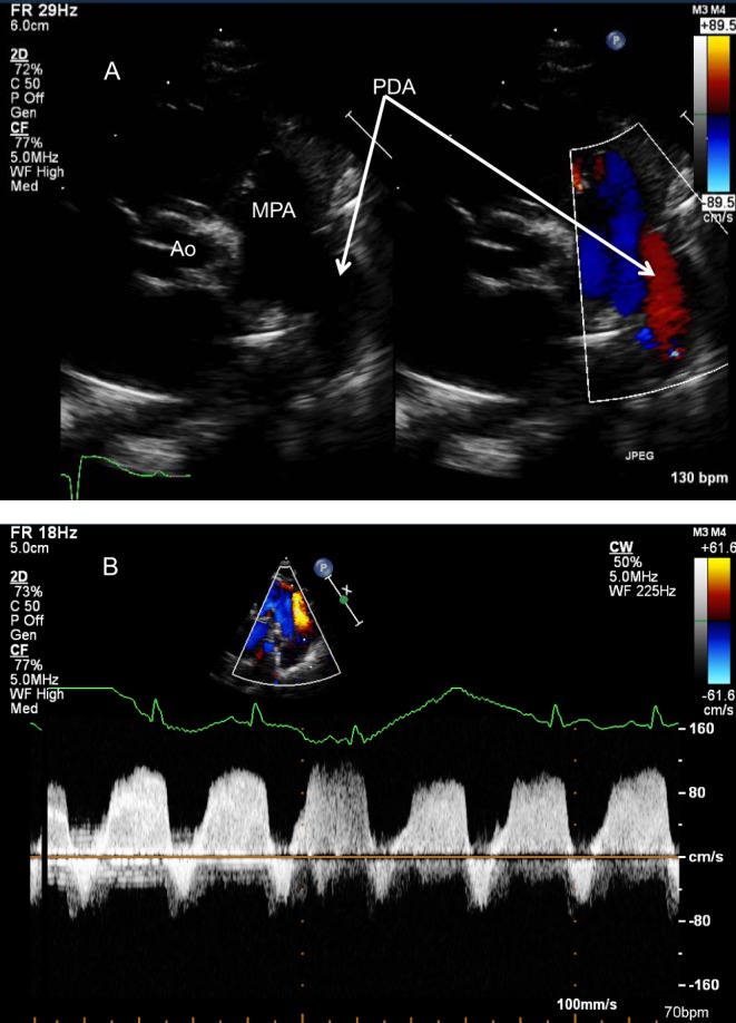
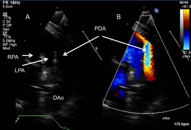
This review article will guide the reader through the basics of echocardiographic assessment of congenital left to right shunts in both paediatric and adult age groups. After reading this article, the reader will understand the pathology and clinical presentation of atrial septal defects (ASDs), ventricular septal defects (VSDs), atrioventricular septal defects (AVSDs) and patent arterial duct. Echocardiography is the mainstay in diagnosis and follow-up assessment of patients with congenital heart disease. This article will therefore describe the echocardiographic appearances of each lesion, and point the reader towards specific features to look for echocardiographically.

Keywords: atrial septal defect; atrioventricular septal defect; echocardiography; patent arterial duct; ventricular septal defect.


Figures

Figure 1
Figure 1
Parasternal short-axis view showing a perimembranous VSD (*) partially covered by aneurysmal tricuspid valve tissue in 2D (A) and colour Doppler (B). (C) Shows the Continuous Doppler trace across the defect. The Bernoulli equation allows the calculation of the pressure gradient (PG) between the right and left ventricles, in this case PG = 4 × 4.72 = 88.36 mmHg. The left ventricular systolic pressure is assumed to be equal to the cuff-measured systolic pressure provided, there is no left ventricular outflow tract obstruction. In this case, the systolic blood pressure of the patient was 115 mmHg. The right ventricular pressure will be the difference of the LV systolic pressure and the PG between the two ventricles = 115−88.36 = 26.64 mmHg.
Figure 2
Figure 2
Different types of interatrial communications.
Figure 3
Figure 3
Subcostal four-chamber view shows a large ostium secundum ASD on 2D (A) and colour Doppler (B).
Figure 4
Figure 4
Superior sinus venosus defect (*) demonstrated from subcostal sagittal view on 2D (A) and Colour Doppler (B).
Figure 5
Figure 5
Subcostal view showing a primum atrial septal defect (*). The bridging leaflets of the common valve are attached to the crest of the septum (**) allowing shunt only at the atrial level. This is a partial AVSD.
Figure 6
Figure 6
Dilated right atrium and right ventricle in an adult with a large secundum ASD (*).
Figure 7
Figure 7
Parasternal short-axis view on 2D (A) and colour Doppler (B) showing a large secundum ASD (*). Note the dilated right atrium (RA).
Figure 8
Figure 8
Four-chamber TOE view (mid-oesophageal, 0°) on 2D (A) and colour Doppler (B). The maximum dimension of the ASD is measured and its relationship with the AV valves assessed.
Figure 9
Figure 9
Transoesophageal view (mid-oesophageal, approximately 45°) of a large ASD on 2D (A) and colour Doppler (B). The aortic rim is deficient.
Figure 10
Figure 10
Large atrial shunt secondary to multiple defects seen from the bicaval TOE view (mid-oesophageal, 90°) on 2D (A) and colour Doppler (B).
Figure 11
Figure 11
Assessment of ASD (*) for device closure. The IVC rim is seen from the transgastric bicaval view (116°) on 2D (A) and colour Doppler (B).
Figure 12
Figure 12
TOE 3D view of the atrial septum from the RA side. The ASD is noted with *.
Figure 13
Figure 13
TOE mid-oesophageal view at 0° showing an ASD device in situ following percutaneous ASD closure.
Figure 14
Figure 14
A perimembranous VSD is shown in the PSAX TTE view in 2D (A) and colour Doppler (B). Note the tricuspid-aortic valve fibrous continuity.
Figure 15
Figure 15
Perimembranous VSD partially covered by tricuspid valve tissue.
Figure 16
Figure 16
Large muscular VSD (*). The LA and LV are significantly dilated.
Figure 17
Figure 17
Parasternal short-axis view showing a doubly committed VSD (*). The right coronary cusp of the aortic valve has a mildly elongated appearance as it is prolapsing through the defect, partially covering it. The aortic and pulmonary valves are in fibrous continuity.
Figure 18
Figure 18
Large malalignment VSD (*) as seen from the apical four-chamber view with anterior angulation to open the left ventricular outflow tract (A) and parasternal long axis view (B). The outlet septum is posteriorly deviated (arrow) creating a very narrow outflow tract. This newborn also had a small, bicuspid aortic valve and interruption of the aortic arch.
Figure 19
Figure 19
TOE in a patient with previous surgical closure of a large VSD (* in image A is in the position of the patch) with a fenestration allowing a residual shunt as shown with colour Doppler (arrow in image B). (C) Post percutaneous device closure of the defect (**).
Figure 20
Figure 20
(A) Normal arrangement of atrioventricular and semilunar valves. In the normal heart the aortic and mitral valves are in fibrous continuity. (B) Common AV valve instead of normal Tricuspid and Mitral valves in AVSD. Note the ‘unwedged’ position of the Aorta in the AVSD setting. The Aorta maintains its fibrous continuity with the common AV valve.
Figure 21
Figure 21
Subcostal left anterior oblique view of an infant with AVSD showing the ‘goose-neck deformity’ of the left ventricular outflow tract.
Figure 22
Figure 22
AVSD with large atrial and ventricular components seen from the apical four-chamber view on 2D (A). Colour Doppler (B) shows a mild degree of central AV valve regurgitation.
Figure 23
Figure 23
Patient several years post repair of AVSD with significant subaortic obstruction. (A) PLAX reveals a subaortic membrane (*) but colour Doppler (B) demonstrates presence of turbulence below the level of the membrane. A 3D volume was acquired from the PLAX window. MPR showed chordal attachments to the LVOT (**) in addition to the subaortic membrane (*) (C and F). The excursion of the aortic leaflets is limited secondary to the membrane (D).
Figure 24
Figure 24
(A) AVSD with a degree of unbalance to the left. (B) The common valve is seen from the subcostal LAO view and the area of the left AV valve to the common AV valve is calculated (AV valve index). (C and D) The inflows are assessed on colour Doppler. Note the smaller size of the LV inflow in comparison to that on the right.
Figure 25
Figure 25
TOE in an adult with primum ASD (*). Colour Doppler interrogation did not reveal shunt at the ventricular level.
Figure 26
Figure 26
(A) Parasternal short-axis in a newborn with pulmonary hypertension. There is bidirectional flow across the duct, confirmed on spectral Doppler (B). Note the low velocity flow in keeping with elevated pulmonary arterial pressure.
Figure 27
Figure 27
(A) High parasternal view on 2D shows the bifurcation of the main pulmonary artery and a large PDA with large left to right shunt (B).

References

    1. Hoffman JIE. The global burden of congenital heart disease. Cardiovascular Journal of Africa 2013. 24 141–145. (10.5830/CVJA-2013-028) - DOI - PMC - PubMed
    1. Levin AR, Spach MS, Boineau JP, Canent RV, Jr, Capp MP, Jewett PH. Atrial pressure-flow dynamics in atrial septal defects (secundum type). Circulation 1968. 37 476–488. (10.1161/01.CIR.37.4.476) - DOI - PubMed
    1. Lopez L, Colan SD, Frommelt PC, Ensing GJ, Kendall K, Younoszai AK, Lai WW, Geva T. Recommendations for quantification methods during the performance of a pediatric echocardiogram: a report from the pediatric measurements writing group of the American Society of Echocardiography Pediatric and Congenital Heart Disease Council. Journal of the American Society of Echocardiography 2010. 23 465–495. (10.1016/j.echo.2010.03.019) - DOI - PubMed
    1. Rudski LG, Lai WW, Afilalo J, Hua L, Handschumacher MD, Chandrasekaran K, Solomon SD, Louie EK, Schiller NB. Guidelines for the echocardiographic assessment of the right heart in adults: a report from the American Society of Echocardiography. Journal of the American Society of Echocardiography 2010. 23 685–713. (10.1016/j.echo.2010.05.010) - DOI - PubMed
    1. Helbing WA, Bosch HG, Maliepaard C, Rebergen SA, van der Geest RJ, Hansen B, Ottenkamp J, Reiber JH, de Roos A. Comparison of echocardiographic methods with magnetic resonance imaging for assessment of right ventricular function in children. American Journal of Cardiology 1995. 76 589–594. (10.1016/S0002-9149(99)80161-1) - DOI - PubMed

LinkOut - more resources