. 2018 Apr 10:275:53-66.
doi: 10.1016/j.jconrel.2018.02.012. Epub 2018 Feb 9.

Macromolecular prodrugs of ribavirin: Polymer backbone defines blood safety, drug release, and efficacy of anti-inflammatory effects

Affiliations

Macromolecular prodrugs of ribavirin: Polymer backbone defines blood safety, drug release, and efficacy of anti-inflammatory effects

Kaja Zuwala et al. J Control Release. .

Abstract

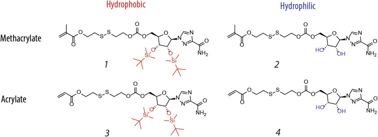
Macromolecular (pro)drugs hold much promise as broad-spectrum antiviral agents as either microbicides or carriers for intracellular delivery of antiviral drugs. Intriguing opportunity exists in combining the two modes of antiviral activity in the same polymer structure such that the same polymer acts as a microbicide and also serves to deliver the conjugated drug (ribavirin) into the cells. We explore this opportunity in detail and focus on the polymer backbone as a decisive constituent of such formulations. Fourteen polyanions (polycarboxylates, polyphosphates and polyphosphonates, and polysulfonates) were analyzed for blood pro/anti coagulation effects, albumin binding and albumin aggregation, inhibitory activity on polymerases, cytotoxicity, and anti-inflammatory activity in stimulated macrophages. Ribavirin containing monomers were designed to accommodate the synthesis of macromolecular prodrugs with disulfide-exchange triggered drug release. Kinetics of drug release was fast in all cases however enhanced hydrophobicity of the polymer significantly slowed release of ribavirin. Results of this study present a comprehensive view on polyanions as backbone for macromolecular prodrugs of ribavirin as broad-spectrum antiviral agents.

Keywords: Albumin; Antiviral; Drug release; Macrmolecular prodrug.


Figures

Unlabelled Image
Graphical abstract
Fig. 1
Fig. 1
Anionic monomers (carboxylates, phosphates and phosphonates, and sulfonates) and ribavirin used in this study for the synthesis of polyanions and macromolecular prodrugs as broad-spectrum antiviral agents. Macromolecular prodrugs were synthesized for (meth)acrylates and (meth)acrylamides only (not for VBZ, SVBS, VSA, VPA).
Fig. 2
Fig. 2
Four RBV prodrug monomers used for copolymerization with the diverse set of anion containing monomers. For details on monomer syntheses and polymerization conditions, see Experimental Section.
Fig. 3
Fig. 3
Blood coagulation time (measured as aPTT) in the presence of polyanions administered at concentration 100 or 10 mg/L. The aPTT was measured for up to 150 s and after that time the sample was considered a strong anticoagulant and its blood coagulation time was set to 150 s. Data for carboxylates are reproduced from Ref. [55]. Dash line indicates the normal blood coagulation time in these experimental conditions and readout. The results are an average of four independent experiments (N = 4; two donors in each experiment) and presented as average ± SD. Statistical significance relative to control was evaluated by a one-way ANOVA with Dunnett's multicomparison post-hoc analysis. *p ≤ 0.05; **p ≤ 0.01; ***p ≤ 0.001.
Fig. 4
Fig. 4
Polyanions and macromolecular prodrugs exhibit structure-dependent binding to albumin via Sudlow I/II sites. Interaction with albumin was quantified using fluorescent probe displacement assay using dansyl asparagine and dansyl sarcosine as probes for Sudlow I and Sudlow II sites, respectively. Fluorescence intensities of the probe in the presence of polyanions were normalized to that in control experiments. 100% probe fluorescence indicates no probe displacement and 0% probe fluorescence implies quantitative displacement and indicates strong interaction between the polymer and albumin. Results are presented as average of three independent experiments ± SD. Statistical significance compared to the control samples was evaluated via one-way ANOVA with Dunnett's multicomparison post-hoc analysis. *p ≤ 0.05; **p ≤ 0.01; ***p ≤ 0.001.
Fig. 5
Fig. 5
Size exclusion chromatography elution profiles for equimolar mixtures of albumin with PEAA, PPAA and PAMPS illustrating strong protein aggregation in the presence of PPAA and minor if any interaction of albumin with PAMPS. For PEAA, protein elution profile shifts to shorter elution times indicating an increased hydrodynamic radius of the solute as a result of the polymer-protein interaction.
Fig. 6
Fig. 6
(A) Schematic illustration of the engineered drug release through the scission of the disulfide bond and the ensuing spontaneous cyclization of the self-immolative linker (B). Illustration of the 1H NMR-based method to monitor and quantify release of RBV from the macromolecular prodrug (PAPA-RBV) through integration of peaks corresponding to bond vs release drug. The proton used for integration is marked in red in panel (A). (For interpretation of the references to colour in this figure legend, the reader is referred to the web version of this article.)
Fig. 7
Fig. 7
Kinetics of drug release for PABA-RBV established using the 1H NMR method. Presented results are an average of three independent experiments ± SD.
Fig. 8
Fig. 8
Drug release analyzed by 1H NMR at 2 h showed a significant decrease in release depending on the hydrophobicity of the carrier in the row of carboxylates (PMAA to PEAA to PPAA), as well as a significantly slower release from the methacrylamide based phosphonate relative to the acrylamide based phosphonate. All data are displayed as mean ± standard deviation from three independent experiments. For comparison a one-way ANOVA (analysis of variance) was performed, followed by a Tukey's post hoc test. ***: P ≤ .001.
Fig. 9
Fig. 9
Activity of the polymerase enzyme in the presence of polyanions (1 or 10 mg/L in the reaction mixture) expressed in % of de novo synthesized nucleic acid relative to the un-inhibited polymerase reaction. Statistical significance compared to control was evaluated via a one-way ANOVA with Dunnett's multicomparison post-hoc analysis. *p ≤ 0.05; **p ≤ 0.01; ***p ≤ 0.001.
Fig. 10
Fig. 10
Concentration-dependent inhibition of inflammation by polyanions and polyanion-RBV conjugates in LPS-stimulated Raw 264.7 macrophages with corresponding cytotoxicity. Raw 264.7 macrophages were stimulated with 1 mg/L LPS and inflammation was measured by quantifying NO via the Griess assay. The concentrations of the polymer which significantly inhibited inflammation without inducing significant toxicity were determined using one-way ANOVA and were marked on the chart with red colour (p < 0.05 *). The results are an average of four independent experiments n = 4 ± SD. Statistical significance of results compared to control was evaluated via a one-way ANOVA with Dunnett's multicomparison post-hoc analysis. (For interpretation of the references to colour in this figure legend, the reader is referred to the web version of this article.)
Fig. 11
Fig. 11
Confocal microscopy of Raw 264.7 macrophages incubated with 40 mg/L FITC labelled polyanion. Cells are further stained with DAPI and Concanavalin A Alexa Fluor® 633 to visualize cell nuclei and cytoplasmic membrane. First three rows present emission from the three fluorochromes, and the bottom row is a merged image. Scale bar 20 μm.
Fig. 12
Fig. 12
Competition assay for stimulation of macrophages with LPS in the presence of PEAA. Raw 264.7 macrophages were preincubated with media or with 50 mg/L PEAA and then stimulated with increasing concentrations of LPS. Inflammation was measured by quantifying NO via the Griess assay. The results are an average of three independent experiments n = 3 ± SD. Lower standard error bars were omitted for clarity. The statistical difference in inflammation between PEAA and non-PEAA treated cells was compared using unpaired Student's t-test. p < 0.01**.

Similar articles

Cited by

References

    1. Marston H.D., Folkers G.K., Morens D.M. Emerging viral diseases: confronting threats with new technologies. Sci. Transl. Med. 2014;6:253ps210. - PubMed
    1. Lozano R., Naghavi M., Foreman K. Global and regional mortality from 235 causes of death for 20 age groups in 1990 and 2010: a systematic analysis for the Global Burden of Disease Study. Lancet. 2010;380:2095–2128. - PMC - PubMed
    1. De Clercq E. The design of drugs for HIV and HCV. Nat. Rev. Drug Discov. 2007;6:1001–1018. - PubMed
    1. De Clercq E., Li G. Approved antiviral drugs over the past 50 years. Clin. Microbiol. Rev. 2016;29:695–747. - PMC - PubMed
    1. Bekerman E., Einav S. Combating emerging viral threats. Science. 2015;348:282–283. - PMC - PubMed

Publication types