. 2018 Sep 1:387:178-190.
doi: 10.1016/j.neuroscience.2018.01.047. Epub 2018 Feb 10.

The Changing Sensory and Sympathetic Innervation of the Young, Adult and Aging Mouse Femur

Affiliations

The Changing Sensory and Sympathetic Innervation of the Young, Adult and Aging Mouse Femur

Stephane R Chartier et al. Neuroscience. .

Abstract

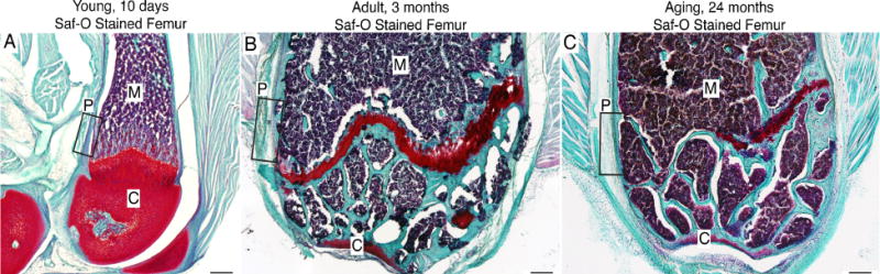
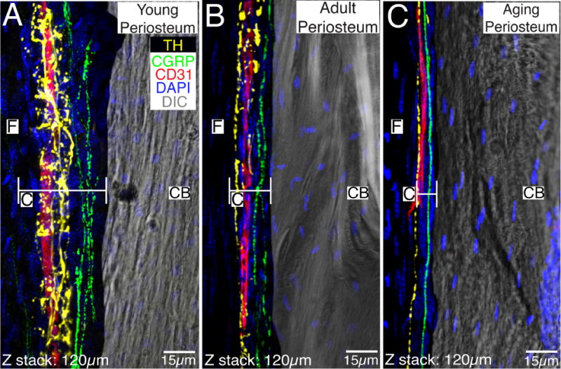
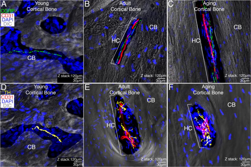
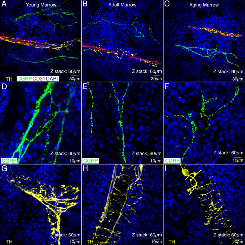
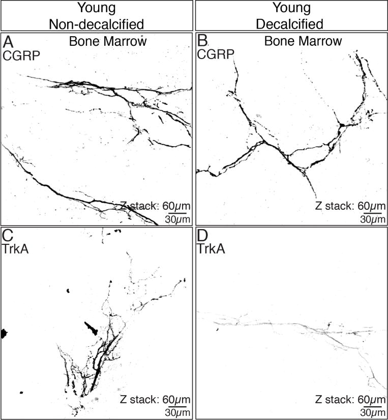
Although bone is continually being remodeled and ultimately declines with aging, little is known whether similar changes occur in the sensory and sympathetic nerve fibers that innervate bone. Here, immunohistochemistry and confocal microscopy were used to examine changes in the sensory and sympathetic nerve fibers that innervate the young (10 days post-partum), adult (3 months) and aging (24 months) C57Bl/6 mouse femur. In all three ages examined, the periosteum was the most densely innervated bone compartment. With aging, the total number of sensory and sympathetic nerve fibers clearly declines as the cambium layer of the periosteum dramatically thins. Yet even in the aging femur, there remains a dense sensory and sympathetic innervation of the periosteum. In cortical bone, sensory and sympathetic nerve fibers are largely confined to vascularized Haversian canals and while there is no significant decline in the density of sensory fibers, there was a 75% reduction in sympathetic nerve fibers in the aging vs. adult cortical bone. In contrast, in the bone marrow the overall density/unit area of both sensory and sympathetic nerve fibers appeared to remain largely unchanged across the lifespan. The preferential preservation of sensory nerve fibers suggests that even as bone itself undergoes a marked decline with age, the nociceptors that detect injury and signal skeletal pain remain relatively intact.

Keywords: genetic disorders; geriatric; nociceptors; pediatric; skeletal.


Conflict of interest statement

All other authors report no conflict of interest.

Figures

Figure 1
Figure 1. Tissue sections of the distal head of the young, adult and aging mouse femur stained with Safranin O
Images are light field photomicrographs of 20μm thick Safranin O (Saf-O) stained sections obtained from (A) 10 day old, (B) 3 month old and (C) 24 month old mouse femur. In these images cartilage is deep red, mineralized bone teal-blue and hemopoietic cells in bone marrow purple. In the present study, we focused on the distal end of the femur although a similar organization and age related changes were also observed in other parts of the femur. The boxes within A, B & C show where the confocal microscopy images in Figures 2–5 were acquired. Abbreviations: M, marrow; C, cartilage; P, periosteum.
Figure 2
Figure 2. Localization of CD31+ blood vessels, CGRP+ primary afferent sensory nerve fibers and TH+ sympathetic nerve fibers in the periosteum of the young, adult and aging mouse femur
In images A, B and C, the periosteum and its fibrous layer (F) are in the left half of the image and the cortical bone (CB) is in the right half of the image. Note that the great majority of blood vessels and nerve fibers are present in the cambium (C) and not the fibrous (F) layer, the cambium undergoes a dramatic thinning with age, and the TH+ sympathetic nerve fibers have a distinct morphology and closer relationship to CD31+ blood vessels as compared to the CGRP+ sensory nerve fibers. Also note that even in the aging periosteum there remains CD31+ blood vessels, TH+ sympathetic nerve fibers, and CGRP+ sensory nerve fibers. In these confocal images the endothelial cells are labeled with an antibody raised against 140 kD glycoprotein known as platelet endothelial cell adhesion molecule which is also known as CD31 (red), sympathetic nerve fibers are labeled by an antibody raised against tyrosine hydroxylase (TH, yellow), and sensory nerve fibers are labeled with an antibody raised against calcitonin gene related peptide (CGRP, green). The nuclei of all cells are stained with DAPI (blue) and the image of one of the sections obtained with differential interference contrast (DIC) microscopy is included to provide orientation. Confocal images in A, B and C were obtained from two serially adjacent tissue sections. The first 60 micron serial section was stained for TH, CD31, and DAPI. The second 60 micron section was stained for CGRP, CD31 and DAPI. These two 60 micron sections were then aligned and stacked on top of each other to generate the 120 micron Z-stack shown in 2A, 2B, and 2C.
Figure 3
Figure 3. Blood vessels, sensory and sympathetic nerve fibers that innervate the cortical bone in the young, adult and aging femur
In these images, the cortical bone (CB) is imaged by differential interfere contrast (DIC) and nuclei within the cortical bone are stained by DAPI (blue). In the young animal, the CB is largely cartilaginous with some woven bone and there are not any well-formed Haversian canals (HC) or CD31+ blood vessels although occasional CGRP+ sensory fibers (A) and TH+ sympathetic (D) nerve fibers can be observed. With aging, both the adult (B&E) and aging (C&F) cortical bone of the femur becomes mineralized and nearly all sensory or sympathetic nerve fibers in the adult and aging cortical bone are present in Haversian canals most of which are vascularized by CD31+ blood vessels. With age, the number of CD31+ Haversian canals declines, as does the percent of CD31+ Haversian canals that contain TH+ sympathetic nerve fibers. A similar decline was not observed in the percent of CD31+ Haversian canals innervated by CGRP+ sensory nerve fibers. It should be noted that we did not observe any sensory or sympathetic nerve fibers in the young, adult or aging subchondral bone. All confocal images are composed of 2 serially adjacent 60 micron sections = total z-stack of 120 microns.
Figure 4
Figure 4. Sensory fibers, sympathetic fibers and blood vessels in the bone marrow of the young, adult and aging mouse femur
In these images, the sympathetic nerve fibers (yellow) are labeled by an antibody raised against tyrosine hydroxylase (TH), the sensory nerve fibers (green) are labeled with an antibody raised against calcitonin gene related peptide (CGRP) and the endothelial cells are labeled with an antibody raised against 140 kD glycoprotein know as platelet endothelial cell adhesion molecule which is also known as CD31 (red). Note that the TH+ sympathetic fibers in the bone marrow have a characteristic “corkscrew shaped” appearance (A, B, C, G, H, I) as they are tightly wrapped around blood vessels. In contrast, CGRP+ sensory nerve fibers have a much more linear appearance (D, E, F) and do not have the same intimate association with blood vessels (A, B, C). Also, note that there is not a significant decline in either the density of the sensory or sympathetic nerve fibers innervation of the bone marrow with age, although sensory nerve fibers do appear to have more branch points with age and the intensity of TH immunoreactivity in the sympathetic fibers appears to decline with age (i.e. compare Figs. G vs. H or I). Confocal images in A, B and C were obtained from two serially adjacent tissue sections. The first 30 micron serial section was stained for TH, CD31, and DAPI. The second 30 micron serial section was stained for CGRP, CD31, and DAPI. These two 30 micron sections were then aligned and stacked on top of each other to generate the 60 micron Z-stack shown in A, B, and C. This figure shows the relationship of CGRP+ sensory, TH+ sympathetic, and CD31+ blood vessels in the young, adult and aging bone marrow, respectively. As one can see in all of these images, the TH+ sympathetic nerve fibers have a distinct morphology and closer relationship to CD31+ blood vessels as compared to the CGRP+ sensory nerve fibers. In (D, E, and F) single 60 micron sections were stained for CGRP and DAPI and in G, H, and I single 60 micron sections were stained with TH and DAPI.
Figure 5
Figure 5. The process of decalcification significantly impacts the ability to detect TrkA but not CGRP immunoreactivity in bone
In the non-decalcified young femur, robust staining of both CGRP (A) and TrkA (C) are observed in the bone marrow. However, while robust CGRP immunoreactivity is still detected following decalcification of the young bone (B), TrkA immunoreactivity (D) markedly declines. These data show that the decalcification procedure (which is required to cut tissue sections from most calcified tissues) can have a marked and negative impact on some but not all antigens. These confocal images were obtained from a single 60 micron section of young bone.

Similar articles

Cited by

References

    1. Allen MR, Hock JM, Burr DB. Periosteum: biology, regulation, and response to osteoporosis therapies. Bone. 2004;35(5):1003–1012. - PubMed
    1. Arnold W. Immunohistochemical investigation of the human inner ear. Limitations and prospects. Acta Otolaryngol. 1988;105(5–6):392–397. - PubMed
    1. Ascano M, Bodmer D, Kuruvilla R. Endocytic trafficking of neurotrophins in neural development. Trends Cell Biol. 2012;22(5):266–273. - PMC - PubMed
    1. Aso K, Ikeuchi M, Izumi M, Sugimura N, Kato T, Ushida T, Tani T. Nociceptive phenotype of dorsal root ganglia neurons innervating the subchondral bone in rat knee joints. Eur J Pain. 2014;18(2):174–181. - PubMed
    1. Bloom AP, Jimenez-Andrade JM, Taylor RN, Castaneda-Corral G, Kaczmarska MJ, Freeman KT, Coughlin KA, Ghilardi JR, Kuskowski MA, Mantyh PW. Breast cancer-induced bone remodeling, skeletal pain, and sprouting of sensory nerve fibers. J Pain. 2011;12(6):698–711. - PMC - PubMed

Publication types