. 2018 Nov;25(1):517-532.
doi: 10.1080/10717544.2018.1435747.

Transferrin and octaarginine modified dual-functional liposomes with improved cancer cell targeting and enhanced intracellular delivery for the treatment of ovarian cancer

Affiliations

Transferrin and octaarginine modified dual-functional liposomes with improved cancer cell targeting and enhanced intracellular delivery for the treatment of ovarian cancer

Pranali Deshpande et al. Drug Deliv. 2018 Nov.

Abstract

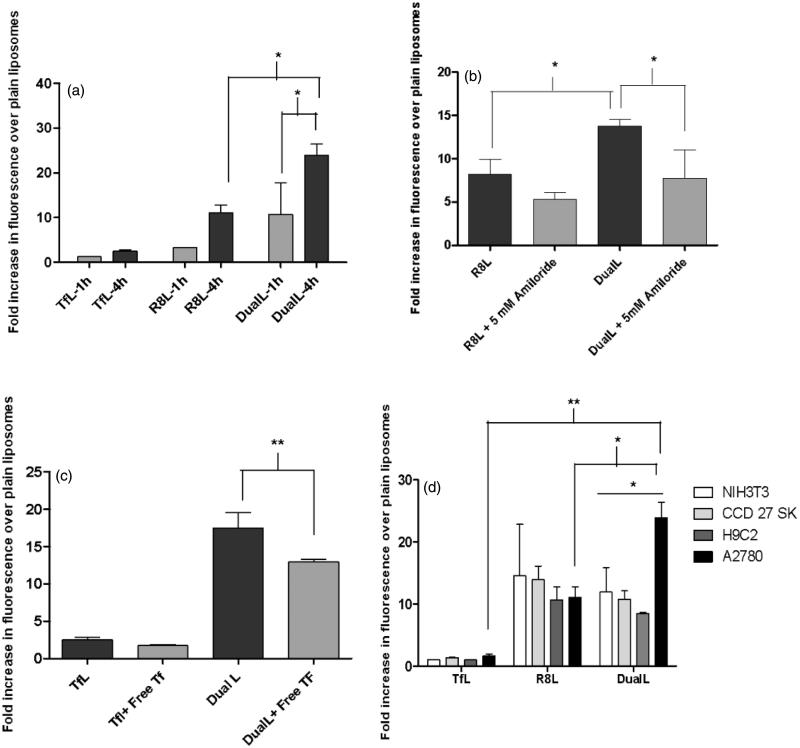
Off-target effects of drugs severely limit cancer therapy. Targeted nanocarriers are promising to enhance the delivery of therapeutics to tumors. Among many approaches for active tumor-targeting, arginine-rich cell penetrating peptides (AR-CPP) and ligands specific to target over-expressed receptors on cancer-cell surfaces, are popular. Earlier, we showed that the attachment of an AR-CPP octaarginine (R8) to the surface of DOXIL® (Doxorubicin encapsulated PEGylated liposomes) improved cytoplasmic and nuclear DOX delivery that enhanced the cytotoxic effect in vitro and improved therapeutic efficacy in vivo. Here, we report on DOX-loaded liposomes, surface-modified with, R8 and transferrin (Tf) (Dual DOX-L), to improve targeting of A2780 ovarian carcinoma cells via the over-expressed transferrin receptors (TfRs) with R8-mediated intracellular DOX delivery. Flow cytometry analysis with fluorescently labeled DualL (without DOX) showed two-fold higher cancer-cell association than other treatments after 4 h treatment. Blocking entry pathways of R8 (macropinocytosis) and Tf (receptor-mediated endocytosis, RME) resulted in a decreased cancer-cell association of DualL. Confocal microscopy confirmed involvement of both entry pathways and cytoplasmic liposome accumulation with nuclear DOX delivery for Dual DOX-L. Dual DOX-L exhibited enhanced cytotoxicity in vitro and was most effective in controlling tumor growth in vivo in an A2780 ovarian xenograft model compared to other treatments. A pilot biodistribution study showed improved DOX accumulation in tumors after Dual DOX-L treatment. All results collectively presented a clear advantage of the R8 and Tf combination to elevate the therapeutic potential of DOX-L by exploiting TfR over-expression imparting specificity followed by endosomal escape and intracellular delivery via R8.

Keywords: Liposomes; TfR over-expression; active targeting; doxorubicin; dual-functional liposomes; octaarginine; ovarian cancer; transferrin.


Figures

Figure 1.
Figure 1.
Transmission electron microscopy images of the modified and unmodified DOX-loaded liposomes. Represents 10,000× magnification, Scale bar, 500 nm.
Figure 2.
Figure 2.
Interaction of rhodamine-labeled dual functional liposomes with A2780 cancer cells analyzed by flow cytometry. (a) Cell association of DualL with cancer cells for 1 h and 4 h treatment periods, followed by analysis by flow cytometry. (b) Effect of a macropinocytosis inhibitor, amiloride (5 mM, 30 min pre-incubation), on R8-mediated cell association for a 4 h liposome treatment period. (c) Evaluation of transferrin receptor-mediated endocytosis by competitive inhibition in presence of free excess Tf (2 mg/ml) in the treatment medium.(d) Comparison of cell association of DualL between NIH3T3, H9C2, CCD 27 SK non-cancer cells and A2780 cancer cells. Cells were incubated with liposomes at a total lipid concentration of 0.1 mg/ml. Results are plotted as fold increase in geometric mean fluorescence over plain liposomes and are mean ± SD, averaged from three separate experiments.*, **, ***, **** indicate p ≤ .05, .01, .001 and .0001, respectively analyzed by one-way ANOVA.
Figure 3.
Figure 3.
Receptor mediated endocytosis and macropinocytosis evaluation to confirm internalization of DualL. (a) A2780 cells were incubated with rhodamine-labeled PL, Tfl, R8L or DualL. Liposomes were added at a total lipid concentration of 0.1 mg/ml for a 4 h treatment period followed by analysis by confocal microscopy. (1) Nuclei stained by Hoechst 33342 at 5 µg/mL for 15 min; (2) Macropinosomes stained by FITC Dextran 70 KDa at 0.35 mg/mL for 30 min before formulation incubation; (3) Endosomes stained with Transferrin-Alexa fluor 680 at 22.5 µg/mL for 15 min; (4) Rhodamine-signal from liposomes; (5) Merged composite picture of all the fluorescence; (6) Co-localization of rhodamine with FITC Dextran (7) Co-localization of rhodamine with Alexa Fluor. Yellow signals in the merged images indicate the co-localization of the red and green, red and pinkish fluorescence represents co-localization of red and gray, respectively. Analysis of fluorescence intensity-colocalization Pearson's coefficient (b) and Mander’s coefficients (c), obtained from the merged pictures (n = 3) from TfL, R8L and DualL-treated cells, by Image J software. The results are mean ± SD averaged from three images of the same treatment. *, **, ***, **** indicate p ≤ .05, .01, .001 and .0001, respectively. Analyzed by Student’s t-test. Scale bar, 25 µm.
Figure 4.
Figure 4.
Intracellular DOX release. A2780 cells were incubated with Dio-labeled PL DOX-L, Tf DOX-L, R8 DOX-L or Dual DOX-L. Liposomes were added at a total lipid concentration of 0.1 mg/ml for 4 h treatment period followed by analysis by confocal microscopy. (a) (1) Nuclei stained by Hoechst 33342 at 5 µg/mL for 15 mins; (2) Dio stain from liposome bilayer; (3) DOX stain; (4) Merged composite picture of all fluorescence; (5) Co-localization of DOX with Hoechst; (6) Co-localization of Dio with DOX; (7) Merged image of Hoechst and Dio. Yellow signals in the merged images indicate the co-localization of the red and green indicating cytoplasmic delivery, and purple fluorescence represents co-localization of red and blue indicating nuclear delivery, respectively. Analysis of fluorescence intensity co-localization (b). Pearson's coefficient and (c). Mander’s coefficient, obtained from the merged pictures (n = 3) from Tf DOX-L, R8 DOX-L and Dual DOX-L-treated cells, by Image J software. The results are mean ± SD averaged from three images of the same treatment. *, **, ***, indicate p ≤ .05, .01 and .001 respectively. Analyzed by Student’s t-test. Scale bar, 10 µm.
Figure 5.
Figure 5.
Effect of dual functional DOX-loaded liposomes on cell death in cancer and non-cancer cells. Assessment of cell viability of A2780 cells treated with DOX-loaded PL DOX-L, Tf DOX-L, R8 DOX-L and Dual DOX-L, at DOX concentration of 0.2—75 µM for 15 min followed by 24 (a) or 48 h (b) incubations. Comparison of cell death in A2780 cancer and NIH3T3, H9C2, CCD27Sk non-cancer cells at 75 µM. Cells treated with DOX-loaded PL DOX-L, Tf DOX-L, R8 DOX-L and Dual DOX-L for 15 min followed by 24 (c) or 48 h (d) incubations. Results obtained as mean ± S.D. from three separate experiments. * indicates p < .05, ** indicates p < .01, *** indicates p < .001analyzed by one-way ANOVA.
Figure 6.
Figure 6.
Evaluation of in vivo therapeutic efficacy of Dual DOX-L. Biodistribution of Dox in mice bearing A2780 tumors. (a) Represents distribution of DOX in major organs. (b) Represents distribution of DOX in tumors across 4 tested groups. A single 10 mg/kg i.v. tail injection of PL DOX-L, Tf DOX-L, R8 DOX-L and Dual DOX-L was administered. After 10 h the mice were sacrificed and major organs and tumors were collected. N = 2 animals per group ± SD. Analyzed by one-way ANOVA where *p < 0.05. Effect of i.v. administration of liposomes on tumor growth in nude mice bearing A2780 tumors. Treatment groups were HBS (control), Free DOX, PL DOX-L, Tf DOX-L, R8 DOX-L, and Dual DOX-L. Arrows indicate day of treatment with the formulations given after tumors reached 50–150 mm3. (n = 4, mean ± SEM). Calculated from day 0 to day 27 of treatment. (c) Represents a line graph showing the trend of tumor growth. (d) Represents a column graph of tumor volumes for all groups. (e) A2780 tumor weights at the end of the study. Treatment was stopped and tumors were excised when the average tumor volume in the control group reached 1000 mm3. Represented as mean ± SEM. *, **, ***, **** indicate p ≤ .05, .01, .001 and .0001, respectively analyzed by Student’s t-test.

References

    1. Allen TM, Sapra P, Moase E. (2002). Use of the post-insertion method for the formation of ligand-coupled liposomes. Cell Mol Biol Lett 7:889–94. - PubMed
    1. Apte A, Koren E, Koshkaryev A, Torchilin VP. (2014). Doxorubicin in TAT peptide-modified multifunctional immunoliposomes demonstrates increased activity against both drug-sensitive and drug-resistant ovarian cancer models. Cancer Biol Ther 15:69–80. - PMC - PubMed
    1. Aslan B, Ozpolat B, Sood AK, Lopez-Berestein G. (2013). Nanotechnology in cancer therapy. J Drug Target 21:904–13. - PMC - PubMed
    1. Bajelan E, Haeri A, Vali AM, et al. (2012). Co-delivery of doxorubicin and PSC 833 (Valspodar) by stealth nanoliposomes for efficient overcoming of multidrug resistance. J Pharm Pharm Sci 15:568–82. - PubMed
    1. Biswas S, Deshpande PP, Perche F, et al. (2013). Octa-arginine-modified pegylated liposomal doxorubicin: an effective treatment strategy for non-small cell lung cancer. Cancer Lett 335:191–200. - PMC - PubMed

MeSH terms