. 2018 Feb 12;37(1):24.
doi: 10.1186/s13046-018-0672-z.

SMURF1 facilitates estrogen receptor ɑ signaling in breast cancer cells

Affiliations

SMURF1 facilitates estrogen receptor ɑ signaling in breast cancer cells

Huijie Yang et al. J Exp Clin Cancer Res. .

Abstract

Background: Estrogen receptor alpha (ER alpha) is expressed in the majority of breast cancers and promotes estrogen-dependent cancer progression. ER alpha positive breast cancer can be well controlled by ER alpha modulators, such as tamoxifen. However, tamoxifen resistance is commonly observed by altered ER alpha signaling. Thus, further understanding of the molecular mechanisms, which regulates ER alpha signaling, is important to improve breast cancer therapy.

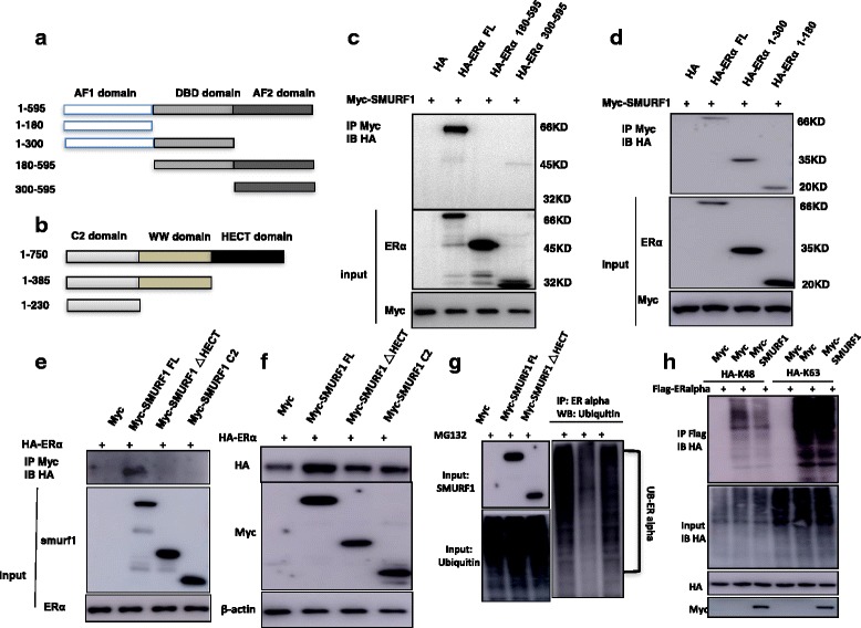
Methods: SMURF1 and ER alpha protein expression levels were measured by western blot, while the ER alpha target genes were measured by real-time PCR. WST-1 assay was used to measure cell viability; the xeno-graft tumor model were used for in vivo study. RNA sequencing was analyzed by Ingenuity Pathway Analysis. Identification of ER alpha signaling was accomplished with luciferase assays, real-time RT-PCR and Western blotting. Protein stability assay and ubiquitin assay was used to detect ER alpha protein degradation. Immuno-precipitation based assays were used to detect the interaction domain between ER alpha and SMURF1. The ubiquitin-based Immuno-precipitation based assays were used to detect the specific ubiquitination manner happened on ER alpha.

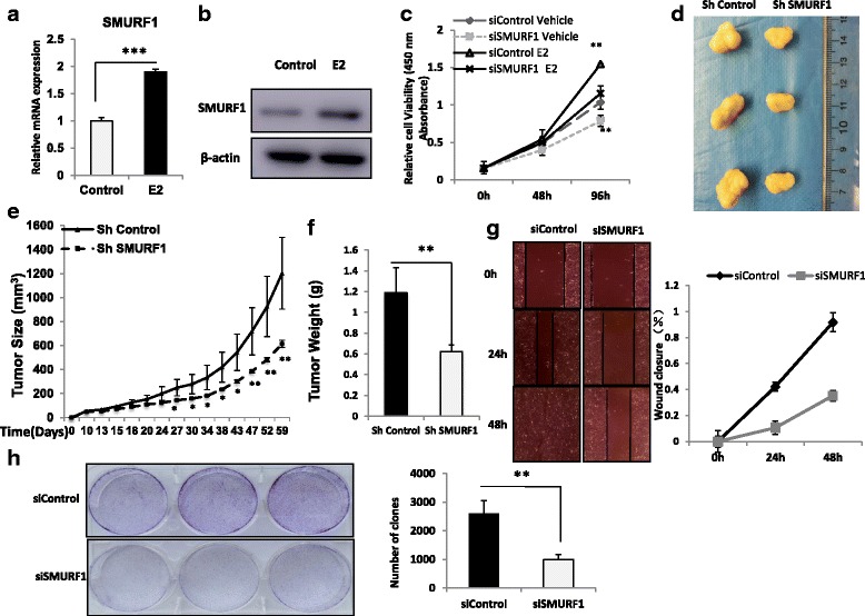
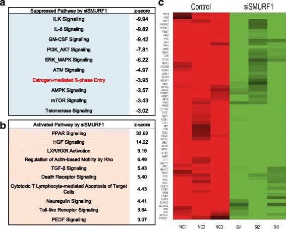
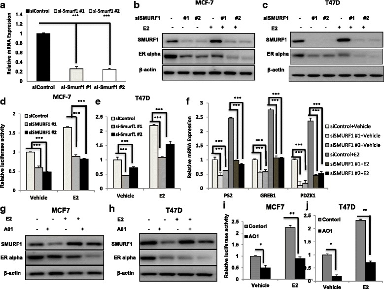
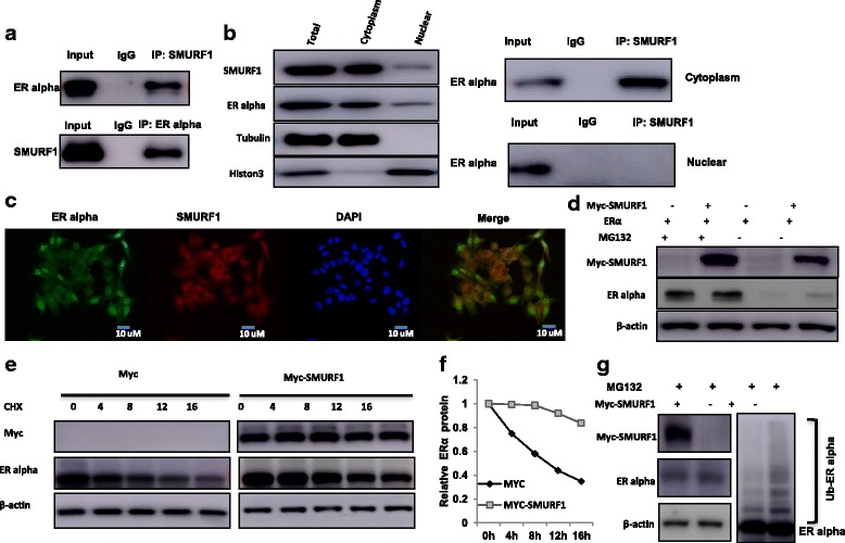
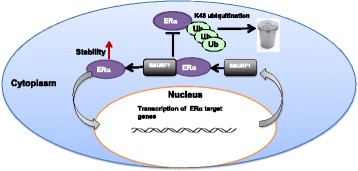
Results: Here, we identify the E3 ligase SMURF1 facilitates ER alpha signaling. We show that depletion SMURF1 decreases ER alpha positive cell proliferation in vitro and in vivo. SMURF1 depletion based RNA-sequence data shows SMURF1 is necessary for ER alpha target gene expression in the transcriptomic scale. Immunoprecipitation indicates that SMURF1 associates with the N-terminal of ER alpha in the cytoplasm via its HECT domain. SMURF1 increases ER alpha stability, possibly by inhibiting K48-specific poly-ubiquitination process on ER alpha protein. Interestingly, SMURF1 expression could be induced via estradiol treatment.

Conclusions: Our study reveals a novel positive feedback between SMURF1 and ER alpha signaling in supporting breast cancer growth. Targeting SMURF1 could be one promising strategy for ER alpha positive breast cancer treatment.

Keywords: Breast cancer; ER alpha; Protein stability; SMURF1; Ubiquitination.


Conflict of interest statement

Ethics approval and consent to participate

This study was reviewed and approved by the Ethical Board at Xinxiang Medical University.

Consent for publication

Not applicable.

Competing interests

The authors’ declare that they have no competing of interest.

Publisher’s Note

Springer Nature remains neutral with regard to jurisdictional claims in published maps and institutional affiliations.

Figures

Fig. 1
Fig. 1
SMURF1 depletion inhibits ER alpha positive breast cancer cell proliferation in vitro and in vivo. a and b Estrogen stimulates the expression of endogenous SMURF1 in breast cancer cells. MCF-7cells are treated with 10 nM estradiol. After 24 h, SMURF1 mRNA and protein levels were determined by Western blot analysis. Actin was used as internal control. Experiments were done in triplicates. *P < 0.05; ** P < 0.01; ***P < 0.001 for SMURF1 mRNA level comparison. c SMURF1 depletion inhibits the cell proliferation in breast cancer cells. MCF-7 cells were transfected with 50 nM SMURF1 siRNA (mix of #1 and #2) or 50 nM control siRNA. After 24 h, the WST assay was used to determine the cellar metabolic activity at indicated time points after transfection. Cells are treated for indicated times with 10 nM E2 or vehicle. Experiments were done in triplicates. *P < 0.05; ** P < 0.01; ***P < 0.001 for cell growth comparison. d, e and f MCF-7 cells were stably transfected with lentivirus carrying scrambe shRNA or SMURF1 shRNA. Female NOD scid gamma (NSG) mice were estrogen-supplemented by implantation of slow-release 17β-estradiol pellets (0.72 mg/90-d release; Innovative Research of America) 1 day before MCF-7 tumor cell injection into the mammary fat pad (2 × 106 MCF-7 cells suspended in 100ul Matrigel solution). MCF-7 tumor xenografts were measured every 3~5 days and the tumor volume were calculated by length × width2 /2. The mice were sacrificed at 2 month after transplant, and the tumors were weighted. The tumor growth curve, photograph and tumor weight were shown in Figure (d), (e) and (f) respectively. g Wound healing assay of MCF-7 transfected with the indicated siRNA. Quantification of wound closure at the indicated time points. Data are presented as ± SD. **, P < 0.01, ***, P < 0.001 (student’s t-test). h Clone formation assay of MCF-7 cells transfected with indicated siRNA. Quantification of clone formation is shown at the indicated time points. Data are presented as ± SD. **, P < 0.01, ***, P < 0.001 (student’s t-test)
Fig. 2
Fig. 2
SMURF1 depletion decreases the expression of ER alpha target genes in breast cancer cells. a Top 10 signaling pathways significantly decreased by SMURF1 depletion in MCF7 cells. The pathway-enrichment analysis was used by the threshold P < 0.001 and fold change > 2 to derive regulated genes. SMURF1 was depleted by siRNA (mix of siSMURF1 #1 and siSMURF1 #2) or treated with siControl. After 48 h, the whole mRNA was extracted for RNA sequence analysis. The siControl and siSMURF1 were done in triplicates. b Top 10 signaling pathways, which were significantly activated by SMURF1 depletion in MCF7 cells. The pathway-enrichment analysis was used by the threshold P < 0.001 and fold change > 2 to derive regulated genes. The pathway-enrichment analysis was used by the threshold P < 0.001 and fold change > 2 to derive regulated genes. SMURF1 was depleted by siRNA (mix of siSMURF1 #1 and siSMURF1 #2) or treated with siControl. After 48 h, the whole mRNA was extracted for RNA sequence analysis. The siControl and siSMURF1 were done in triplicates. c The heat-map graph shows the ERα regulating genes, which is significantly changed by SMURF1 depletion in MCF-7 cells
Fig. 3
Fig. 3
SMURF1 depletion or inhibition in breast cancer cells decreases ER alpha signaling activity. a SMURF1 depletion effect by two different siRNA oligos. MCF-7 cells are transfected with two independent SMURF1 siRNAs or siControl. After 48 h, SMURF1 mRNA levels are determined by QPCR. 36B4 was used as internal control. *P < 0.05; ** P < 0.01; ***P < 0.001 for SMURF1 mRNA level comparison. b and c SMURF1 depletion effect on ERα protein level by two different siRNA oligos. MCF-7 or T47D cells were transfected with siSMURF1 or siControl. After 48 h, cells were treated with either ethanol or 10 nM estradiol for 6 h. SMURF1 and ERα protein levels were determined by Western blot analysis. Actin was used as internal control. d and e SMURF1 depletion affects ERE-luciferase activity in MCF7 and T47D cells. MCF7 or T47D cells were transfected with siSMURF1 or siControl together with ERE luciferase reporter plasmid. Cells were treated with 10 nM estradiol or vehicle. Luciferase activity was measured 48 h after transfection. Shown are the results from three experiments. *P < 0.05; ** P < 0.01; ***P < 0.001 for luciferase activity comparison. f SMURF1 depletion decreases ERα target genes using two different siRNA oligos. MCF-7 cells were transfected with siSMURF1 or siControl. After 48 h, cells were treated with either ethanol or 10 nM estradiol for 6 h. Total RNA was prepared and the expression of the endogenous ERα target genes, PS2, GREB1, and PDZK1 were determined by qPCR. Shown are the results from three experiments. *P < 0.05; ** P < 0.01; ***P < 0.001 for target gene expression comparison. g and h SMURF1 inhibitor A01 decreases ER alpha protein in breast cancer cells. MCF-7 and T47D cells were both treated with 10 nM E2 or vehicle for 24 h and then continue with 10 uM A01 or vehicle 12 h,ER alpha and SMURF1 levels were determined by western blot analysis. β-actin was used as internal control. i and j SMURF1 inhibitor A1 decrease ERE-luciferase activity in MCF7 and T47D cells. MCF7 or T47D cells were transfected with ERE luciferase reporter plasmid. Cells were both treated with 10 nM E2 or vehicle for 24 h and then continue with 10 uM A01 or vehicle for 12 h. Luciferase activity was measured. Shown are the results from three experiments. *P < 0.05; ** P < 0.01; ***P < 0.001 for luciferase activity comparison
Fig. 4
Fig. 4
SMURF1 associates with ER alpha and increases its stability. a Co-IP assay reveals association between endogenous SMURF1 and ERα in MCF7 cells. MCF-7 cells were harvested with NP-40 lysis buffer. CO-IP was performed using antibody as indicated. b SMURF1 is mainly localized in the cytoplasm and associates with ER alpha in the cytosol. The subcellular protein fractionation kit (Thermo scientific, 78,840) was used for cytoplasm and nuclear separation. Tubulin and Histone-3 were used for cytoplasm and nuclear control. Based on the separation, IP was done by SMURF1 antibody in both the cytosol and nuclear lysis. ER alpha antibody was used to detect the interaction in both the cytosol and nuclear. c Intracellular localization analysis of SMURF1 and ER alpha by immunofluorescence assay. MCF7 cells were cultured in phenol red-free DMEM medium. Intracellular localization of SMURF1 (red) and ER alpha (green) were shown. Nuclei (blue) were stained with 4′,6-diamidino-2-phenylindole (DAPI). d In the presence of the proteasome inhibitor MG132, the stabilization effect of SMURF1 on ER alpha did not further increase ER alpha protein levels. HEK293 cells were transfected with 2 μg ERα plasmid and 0.5 μg Myc-tag or Myc-SMURF1 plasmids. After 24 h, cells were treated with 10 uM MG132/vehicle for 6 h. Cell lysates were prepared for Western blot analysis. The results are representative for three independent experiments. e and f SMURF1 increases ERα half-life in HEK293 cells. HEK293 cells were transfected with HA-ERα plasmid and Myc tag or Myc-SMURF plasmids. After 24 h, cells were treated with 100 μM cycloheximide/vehicle for indicated times. Cell lysates were prepared for Western blot analysis. The results are representative for three independent experiments. The ER alpha relative density was measured by Image J software. g SMURF1 prohibits ERα poly-ubiquitination. HEK293 cells were transfected with 2 μg ERα plasmid and 0.5 μg Myc-tag or Myc-SMURF1 plasmids. After 24 h, cells were treated with 10 uM MG132 or vehicle for 6 h. Cells were directly harvested and Western blot analysis using ERα antibody was used to detect ubiquitinated ERα forms. The predicted molecular weight of polyubiquitinated ERα is indicated
Fig. 5
Fig. 5
SMURF1 interacts with ER alpha AF1 domain throμgh its HECT domain and prohibits ER alpha K48 specific poly-ubiquitination. a ER alpha domain structure and deletion mutants used in the study (Full length, ΔAF1, ΔAF1 + ΔDBD, ΔAF2, ΔAF2 + ΔDBD). b SMURF1 domain structure and deletion mutants used in the study (Full length, ΔHECT, ΔHECT + ΔWW). c and d SMURF1 interacts with ER alpha throμgh its AF1 domain. HEK293 cells were transfected with 2 μg Myc-SMURF1 together with HA-ER alpha full length or mutants (ΔAF1, ΔAF1 + ΔDBD, ΔAF2 and ΔAF2 + ΔDBD). After 24 h, cells were harvested with NP-40 lysis buffer. CO-IP was performed using Myc antibody. The possible interacted ER alpha domains were detected by HA antibody. e HECT domain is required for SMURF1 interaction with ER alpha. HEK293 cells were transfected with 2 μg HA-ER alpha together with Myc-SMURF1 full length or mutants (ΔHECT, ΔHECT + ΔWW). After 24 h, cells were harvested with NP-40 lysis buffer. CO-IP was performed using HA antibody. The possible interacted SMURF domains were detected by Myc antibody. f The HECT domain is necessary for the SMURF1-mediated increase of ER alpha protein level. HEK293 cells were transfected with 2 μg HA-ER alpha together with Myc-SMURF1 full length or mutants (ΔHECT, ΔHECT + ΔWW). After 48 h, whole cell extracts were prepared and the level of ER alpha protein was assayed by western blot analysis. g The HECT domain is necessary for SMURF1 inhibition effect on ER alpha poly-ubiquitination. HEK293 cells were transfected with 2 μg Flag-ER alpha plasmid, 0.5 μg HA-Ub plasmid and 0.5 μg Myc-SMURF1/Myc-SMURF1_delta HECT/Myc-vector plasmids. The cell extracts were immunoprecipitated with ER alpha antibody. The poly-ubiquitinated ER alpha was detected by HA antibody. h SMURF1 decreases K48-linked poly-ubiquitination of ER alpha. HEK293 cells were transfected with 2 μg Flag-ER alpha plasmid, 0.5 μg HA-K48 Ubi/HA-K63 Ubi plasmid and 0.5 μg Myc-SMURF1 plasmids. The cell extracts were immunoprecipitated with HA antibody. The K48 specific poly-ubiquitinated ER alpha or K63 specific poly-ubiquitinated was detected via western blotting analysis
Fig. 6
Fig. 6
The hypothetical model for the positive feedback loop between SMURF1 and ER alpha signaling in breast cancer cells: ER alpha signaling induces the expression of SMURF1, which subsequently stabilizes ER alpha via its HECT domain possibly by inhibiting ER alpha K48-linked polyubiqutination process

References

    1. Ferlay J, Soerjomataram I, Dikshit R, Eser S, Mathers C, Rebelo M, Parkin DM, Forman D, Bray F. Cancer incidence and mortality worldwide: sources, methods and major patterns in GLOBOCAN 2012. Int J Cancer. 2015;136(5):E359–E386. doi: 10.1002/ijc.29210. - DOI - PubMed
    1. Reis-Filho JS, Pusztai L. Gene expression profiling in breast cancer: classification, prognostication, and prediction. Lancet. 2011;378(9805):1812–1823. doi: 10.1016/S0140-6736(11)61539-0. - DOI - PubMed
    1. Kumar R, Zakharov MN, Khan SH, Miki R, Jang H, Toraldo G, Singh R, Bhasin S, Jasuja R. The dynamic structure of the estrogen receptor. J Amino Acids. 2011;2011:812540. doi: 10.4061/2011/812540. - DOI - PMC - PubMed
    1. Lupien M, Eeckhoute J, Meyer CA, Krum SA, Rhodes DR, Liu XS, Brown M. Coactivator function defines the active estrogen receptor alpha cistrome. Mol Cell Biol. 2009;29(12):3413–3423. doi: 10.1128/MCB.00020-09. - DOI - PMC - PubMed
    1. Breast International Group 1-98 Collaborative G. Thurlimann B, Keshaviah A, Coates AS, Mouridsen H, Mauriac L, Forbes JF, Paridaens R, Castiglione-Gertsch M, Gelber RD, et al. A comparison of letrozole and tamoxifen in postmenopausal women with early breast cancer. N Engl J Med. 2005;353(26):2747–2757. doi: 10.1056/NEJMoa052258. - DOI - PubMed

MeSH terms