

Takayasu Arteritis Coexisting with Sclerosing Osteomyelitis
- PMID: 29434141
- PMCID: PMC6064701
- DOI: 10.2169/internalmedicine.0329-17
Takayasu Arteritis Coexisting with Sclerosing Osteomyelitis
Abstract
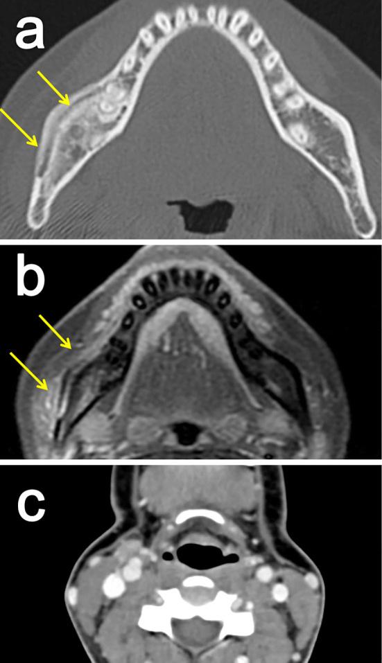
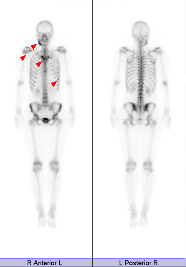
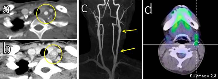
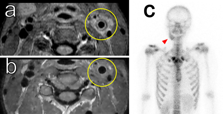
We report a rare case of a 27-year-old woman with Takayasu arteritis (TAK) complicated by diffuse sclerosing osteomyelitis. She first presented with sclerosing osteomyelitis of the right mandible without evidence of arteritis in the carotid arteries. Eight months later, she complained of left neck pain, and imaging studies revealed the presence of arteritis in the left carotid artery. She was diagnosed with TAK, and immunosuppressive treatment was initiated, which was effective for both the arteritis and the osteomyelitis. Osteomyelitis is an important complication of TAK and bone scintigraphy is useful for its detection.
Keywords: Takayasu arteritis; chronic recurrent multifocal osteomyelitis; osteomyelitis; synovitis-acne-pustulosis-hyperostosis-osteitis syndrome.
Figures
References
-
- de Sauza AW, de Carvalho JF. Diagnostic and classification criteria of Takayasu arteritis. J Autoimmun 48-49: 79-83, 2014. - PubMed
-
- Terao C, Yoshifuji H, Ohmura K, et al. Association of Takayasu arteritis with HLA-B 67:01 and two amino acids in HLA-B protein. Rheumatology 52: 1769-74, 2013. - PubMed
-
- Ohta Y, Ohya Y, Fujii K, et al. Inflammatory disease associated with Takayasu artiritis. Angiology 54: 339-344, 2003. - PubMed
-
- Kobak S. Relapsing polychondritis-associated Takayasu's arteritis. Folia Med 51: 49-52, 2009. - PubMed
Publication types
MeSH terms
LinkOut - more resources
Full Text Sources
Other Literature Sources
Miscellaneous