. 2018 Feb 12;8(1):2855.
doi: 10.1038/s41598-018-21184-5.

Early Changes of Articular Cartilage and Subchondral Bone in The DMM Mouse Model of Osteoarthritis

Affiliations

Early Changes of Articular Cartilage and Subchondral Bone in The DMM Mouse Model of Osteoarthritis

Hang Fang et al. Sci Rep. .

Abstract

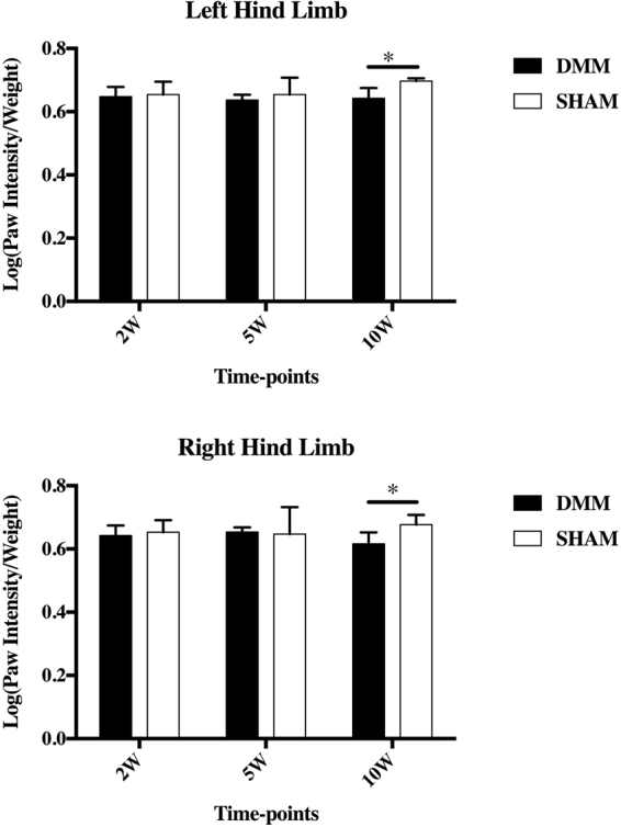
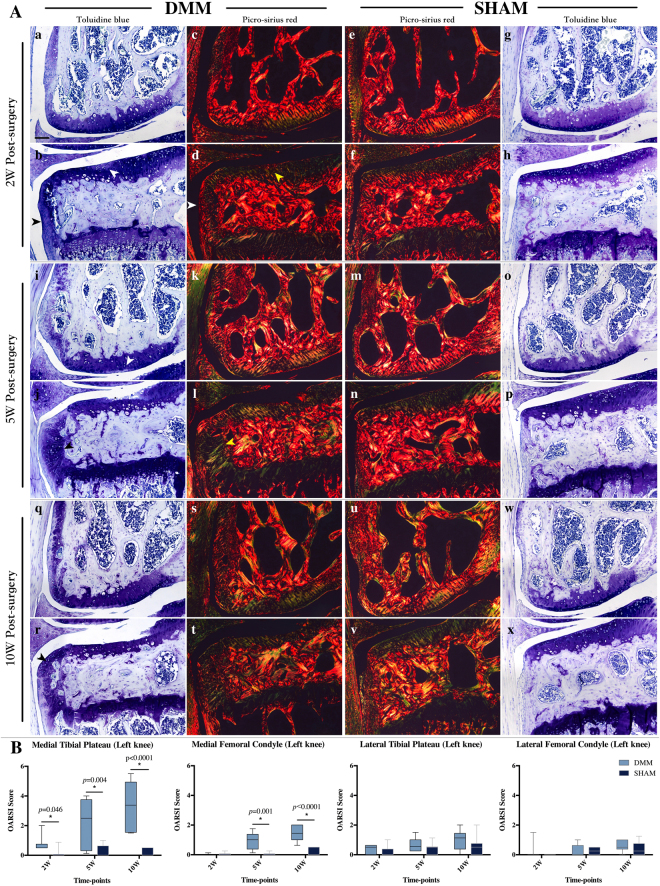
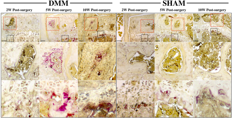
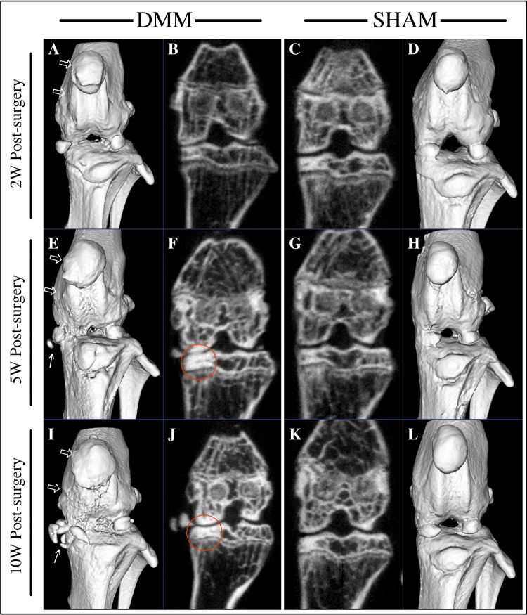
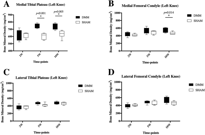
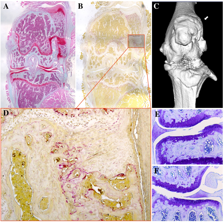
To examine the early changes of articular cartilage and subchondral bone in the DMM mouse model of osteoarthritis, mice were subjected to DMM or SHAM surgery and sacrificed at 2-, 5- and 10-week post-surgery. Catwalk gait analyses, Micro-Computed Tomography, Toluidine Blue, Picrosirius Red and Tartrate-Resistant Acid Phosphatase (TRAP) staining were used to investigate gait patterns, joint morphology, subchondral bone, cartilage, collagen organization and osteoclasts activity, respectively. Results showed OA progressed over 10-week time-course. Gait disparity occurred only at 10-week post-surgery. Osteophyte formed at 2-week post-surgery. BMDs of DMM showed no statistical differences comparing to SHAM at 2 weeks, but BV/TV is much higher in DMM mice. Increased BMD was clearly found at 5- and 10-week post-surgery in DMM mice. TRAP staining showed increased osteoclast activity at the site of osteophyte formation of DMM joints at 5- and 10-week time points. These results showed that subchondral bone turnover might occurred earlier than 2 weeks in this mouse DMM model. Gait disparity only occurred at later stage of OA in DMM mice. Notably, patella dislocation could occur in some of the DMM mice and cause a different pattern of OA in affected knee.


Conflict of interest statement

The authors declare no competing interests.

Figures

Figure 1
Figure 1
Catwalk gait analysis after DMM surgery. Catwalk analyses demonstrated that paw intensity of both hind limbs was lower in DMM mice compared to SHAM at the 10-week time point. DMM surgery was performed in the left hind limb and the right limb was not operated. No statistical significance was detected at the earlier time-points. Error bar stands for the 95% confidence interval.
Figure 2
Figure 2
Histological analyses of joint changes after DMM surgery. (A) Toluidine blue and picrosirius red staining demonstrated that OA progressed from mild to moderate or severe through the 10-week time course on the medial tibial plateau. Regional proteoglycan loss (b, white arrow), chondrocyte clustering and cartilagenous osteophyte formation (b, black arrow) can be detected at 2 weeks post-surgery. Picrosirius red staining showed changes in osteophyte development from 2- to 10-week post-surgery (d,l,t). Scale bar: 200 µm. (B) OARSI scores of the medial tibial plateau of DMM mice were increased at all time-points, while statistically significant increases in the score of the DMM medial femoral condyle was observed at 5- and 10-week time-points (B). No statistical difference in scores was seen on the lateral side of the joint.
Figure 3
Figure 3
Tartrate-resistant acid phosphatase (TRAP) staining of osteoclast activity after DMM surgery. Osteoclast activity in the subchondral bone appeared increased at 5- and 10-week following DMM surgery, mostly within the osteophyte (red rectangle), but not at 2-weeks. The expected staining at the growth plate was observed (black rectangle). Scale bar: 200 µm.
Figure 4
Figure 4
µCT 3D joint reconstruction of DMM-operated mice. Micro-CT analyses demonstrated that joint condition deteriorated through the time course in DMM mice, presenting with uneven bone surface, abnormal patella shape and osteophyte formation (E,I, arrow) around the joint. A coronal plane that represents the middle of the joint showed bone sclerosis in the medial tibial plateau at 5 and 10 weeks post-surgery in DMM mice. (F,J, circle).
Figure 5
Figure 5
Bone mineral density (BMD) of subchondral bone in the DMM model. At 2 weeks pot-surgery, BMDs of the MTP in DMM mice didn’t showed statistical significant differences comparing to their SHAM controls. However, DMM mice showed a larger spread of BMD at 2 weeks post-surgery (A). Clearer differences were detected between DMM and SHAM at both 5 and 10 weeks post-surgery with higher BMDs in DMM mice. (A). BMDs of the MFC were higher in DMM mice 10 weeks after surgery (B). There was no significant differences found in the lateral compartments between DMM and SHAM at all time-points (C,D). Error bar stands for the 95% confidence interval.
Figure 6
Figure 6
Possible subluxation/dislocation of the patella after DMM surgery. Safranin-O staining showed the out-grown osteophyte stained strongly for GAG (A). TRAP staining demonstrated increased number and activity of osteoclasts (B,D). µCT showed out-grown osteophyte formation (C, arrow) and deteriorated joint condition (C). Toluidine Blue staining showed cartilage degeneration on medial (E) and lateral (F) compartments at 10 weeks post-Surgery in DMM mice with patella dislocation.

References

    1. Castañeda S, Roman-Blas JA, Largo R, Herrero-Beaumont G. Subchondral bone as a key target for osteoarthritis treatment. Biochem. Pharmacol. 2012;83:315–323. doi: 10.1016/j.bcp.2011.09.018. - DOI - PubMed
    1. Zhang R, et al. Gene expression analyses of subchondral bone in early experimental osteoarthritis by microarray. PLoS ONE. 2012;7:e32356. doi: 10.1371/journal.pone.0032356. - DOI - PMC - PubMed
    1. Burr DB, Gallant MA. Bone remodelling in osteoarthritis. Nat Rev Rheumatol. 2012;8:665–673. doi: 10.1038/nrrheum.2012.130. - DOI - PubMed
    1. Sanchez C, et al. Subchondral bone osteoblasts induce phenotypic changes in human osteoarthritic chondrocytes. Osteoarthr. Cartil. 2005;13:988–997. doi: 10.1016/j.joca.2005.07.012. - DOI - PubMed
    1. Sanchez C, et al. Osteoblasts from the sclerotic subchondral bone downregulate aggrecan but upregulate metalloproteinases expression by chondrocytes. This effect is mimicked by interleukin-6, -1beta and oncostatin M pre-treated non-sclerotic osteoblasts. Osteoarthr. Cartil. 2005;13:979–987. doi: 10.1016/j.joca.2005.03.008. - DOI - PubMed

Publication types

Grants and funding

LinkOut - more resources