. 2018 Feb 12;8(1):2833.
doi: 10.1038/s41598-018-21324-x.

Murine hematopoietic stem cell reconstitution potential is maintained by osteopontin during aging

Affiliations

Murine hematopoietic stem cell reconstitution potential is maintained by osteopontin during aging

Jin Li et al. Sci Rep. .

Abstract

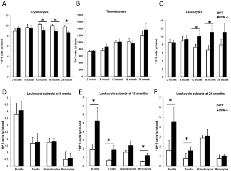
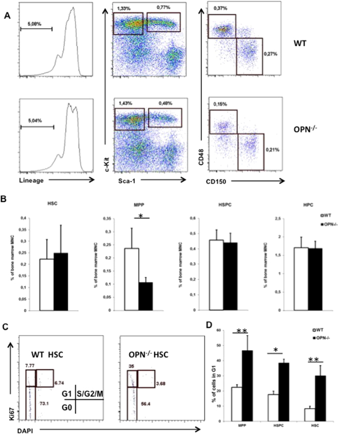
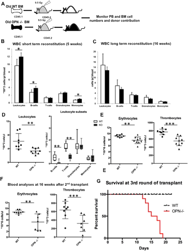
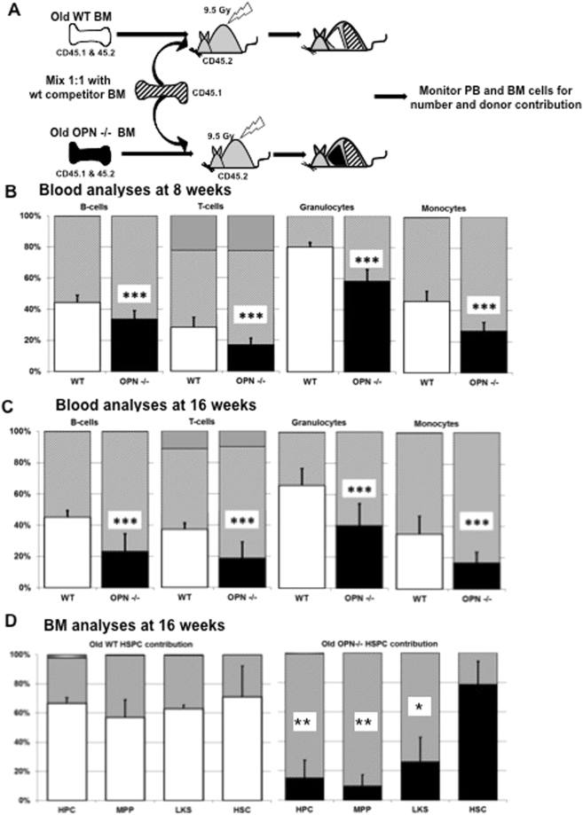
In adult mammals, hematopoietic stem cells (HSCs) reside in the bone marrow and are in part regulated by the bone marrow microenvironment, called the stem cell niche. We have previously identified the bone marrow morphogen osteopontin (OPN), which is abundantly present in the bone marrow extracellular matrix, as a negative regulator of the size of the HSC pool under physiological conditions. Here, we study the impact of OPN on HSC function during aging using an OPN-knockout mouse model. We show that during aging OPN deficiency is associated with an increase in lymphocytes and a decline in erythrocytes in peripheral blood. In a bone marrow transplantation setting, aged OPN-deficient stem cells show reduced reconstitution ability likely due to insufficient differentiation of HSCs into more mature cells. In serial bone marrow transplantation, aged OPN-/- bone marrow cells fail to adequately reconstitute red blood cells and platelets, resulting in severe anemia and thrombocytopenia as well as premature deaths of recipient mice. Thus, OPN has different effects on HSCs in aged and young animals and is particularly important to maintain stem cell function in aging mice.


Conflict of interest statement

The authors declare no competing interests.

Figures

Figure 1
Figure 1
OPN deficiency alters peripheral blood cell counts in aged mice. (AC) Cell counts of erythrocytes, thrombocytes, and leukocytes respectively in young and aged mice. (DF) Cell counts of leukocyte subtypes defined by surface marker expression using flow cytometry in young (D), 18 month (E) and 24 month (F) old mice. Values are the mean ± SEM; n ≥ 12; *P ≤ 0.05.
Figure 2
Figure 2
OPN alters the composition of the primitive HSC compartment. (A) Representative plots from 22-month-old WT and OPN−/− bone marrow cells. (B) Cumulative data of HSPC subpopulations calculated as percentage of total bone marrow mononuclear cells. Values are the mean ± SD; n = 7; *P ≤ 0.05. (C) Representative plots of cell cycle analyses by Ki67 and DAPI gated on HSC population. (D) Cumulative analyses of the proportion of stem and progenitor cells in G1 phase of the cell cycle in WT and OPN−/− mice. Values are the mean ± SD; n = 3; *P ≤ 0.05, **P ≤ 0.01.
Figure 3
Figure 3
OPN deficiency results in loss of serial reconstitution ability in aged animals. (A) Schematic of the transplantation and analysis setting. Bone marrow from CD45.1-bearing WT or OPN-KO mice was transplanted into lethally irradiated CD45.2-bearing recipients and monitored for peripheral blood and bone marrow reconstitution. (B) Short- and (C) long-term analysis of peripheral blood cells at 5 and 16 weeks after transplantation. (D) Total leukocyte count and leukocyte subsets from a secondary bone marrow transplant. (E) Red blood cell and platelet counts in the secondary recipients of aged OPN−/− and WT bone marrow 10 weeks after bone marrow transplant. (F) Analysis of blood cell counts of the same mice 16 weeks after the second round of bone marrow transplant. (G) Kaplan Meier plot indicating survival after third round of bone marrow transplant. Values are the mean ± SD, n ≥ 7, *P < 0.05, **P < 0.01, ***P < 0.001.
Figure 4
Figure 4
Aged OPN-deficient bone marrow cells display reduced reconstitution ability in a competitive transplantation setting. (A) Schematic of the competitive repopulation assay, which was performed to test the ability of aged OPN mutant stem cells to compete against WT HSCs. Analyses of the peripheral blood cells 8 weeks (B) and 16 weeks (C) after transplantation. (D) Further cell subtype analyses of the bone marrow compartment. Values are the mean ± SD, n = 9, *P < 0.05, **P < 0.01, ***P < 0.001.

References

    1. Rando TA. Stem cells, ageing and the quest for immortality. Nature. 2006;441:1080–1086. doi: 10.1038/nature04958. - DOI - PubMed
    1. Rossi DJ, Jamieson CHM, Weissman IL. Stems cells and the pathways to aging and cancer. Cell. 2008;132:681–696. doi: 10.1016/j.cell.2008.01.036. - DOI - PubMed
    1. Pearce DJ, Anjos-Afonso F, Ridler CM, Eddaoudi A, Bonnet D. Age-dependent increase in side population distribution within hematopoiesis: implications for our understanding of the mechanism of aging. Stem cells (Dayton, Ohio) 2007;25:828–835. doi: 10.1634/stemcells.2006-0405. - DOI - PubMed
    1. Rossi DJ, et al. Cell intrinsic alterations underlie hematopoietic stem cell aging. Proceedings of the National Academy of Sciences of the United States of America. 2005;102:9194–9199. doi: 10.1073/pnas.0503280102. - DOI - PMC - PubMed
    1. Beerman I, et al. Functionally distinct hematopoietic stem cells modulate hematopoietic lineage potential during aging by a mechanism of clonal expansion. Proceedings of the National Academy of Sciences of the United States of America. 2010;107:5465–5470. doi: 10.1073/pnas.1000834107. - DOI - PMC - PubMed

Publication types

MeSH terms