. 2018 Mar;25(3):289-296.
doi: 10.1038/s41594-018-0028-6. Epub 2018 Feb 12.

Yeast surface display platform for rapid discovery of conformationally selective nanobodies

Affiliations

Yeast surface display platform for rapid discovery of conformationally selective nanobodies

Conor McMahon et al. Nat Struct Mol Biol. 2018 Mar.

Abstract

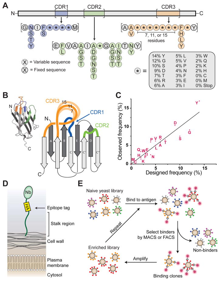
Camelid single-domain antibody fragments ('nanobodies') provide the remarkable specificity of antibodies within a single 15-kDa immunoglobulin VHH domain. This unique feature has enabled applications ranging from use as biochemical tools to therapeutic agents. Nanobodies have emerged as especially useful tools in protein structural biology, facilitating studies of conformationally dynamic proteins such as G-protein-coupled receptors (GPCRs). Nearly all nanobodies available to date have been obtained by animal immunization, a bottleneck restricting many applications of this technology. To solve this problem, we report a fully in vitro platform for nanobody discovery based on yeast surface display. We provide a blueprint for identifying nanobodies, demonstrate the utility of the library by crystallizing a nanobody with its antigen, and most importantly, we utilize the platform to discover conformationally selective nanobodies to two distinct human GPCRs. To facilitate broad deployment of this platform, the library and associated protocols are freely available for nonprofit research.


Figures

Figure 1
Figure 1. Design and construction of synthetic nanobody library
(A) Schematic of synthetic nanobody. Framework regions were fixed in sequence (gray), while portions of the CDR loops were varied (blue, green, and orange, for CDRs 1, 2, and 3, respectively). Partial randomization was achieved with mixed nucleotides to allow up to six possible residues (dotted lines), while highly variable regions were synthesized using a trimer phosphoramidite mixture (asterisks with dotted outlines). (B) Overview of a nanobody showing three-dimensional structure (inset) as well as a cartoon schematic highlighting CDR loops. Synthetic CDR3 sequences used variable lengths of 7, 11, or 15 residues of trimer phosphoramidite mixture. (C) After the library was constructed, amino acid frequencies of diversified positions in CDRs were analyzed by next-generation sequencing (NGS), showing that library frequencies closely matched target values. (D) Schematic of nanobody display on yeast. Nanobody is shown with an HA tag at the carboxy terminus followed by a long flexible stalk which covalently tethers the nanobody to the yeast cell wall. (E) Schematic of the nanobody selection process. Antigen is shown with a fluorescent tag as a glowing red circle. Yeast displaying nanobodies with affinity to antigen are shown being isolated via MACS or FACS, amplified, and undergoing iterative rounds of selection. See also Supplementary Figures 1 and 2, as well as Supplementary Table 1.
Figure 2
Figure 2. Validation of nanobody platform using human serum albumin (HSA) as the target antigen
(A) Histogram of yeast library HSA binding, showing pre-selection (black) and after four rounds (blue). The percentage of HSA-binding cells is indicated. The fraction of the total library composed by Nb.b201 was assessed by deep sequencing, showing progressive enrichment of this clone. (B) Size exclusion chromatography analysis confirmed binding of purified recombinant Nb.b201 to HSA. (C) Crystal structure of nanobody:HSA complex (PDB ID 5VNW), including close-up view of CDR loops interacting with antigen. Hydrogen bonds are shown as dotted lines. (D) Rotated view of the complex (E) Comparison of nanobody structures in the antigen-bound (yellow) and antigen-free state (gray; PDB ID 5VNV), showing conformational change upon antigen binding. Changes are highlighted with arrows, and CDR loops are colored blue, green, and orange for CDRs 1, 2, and 3, respectively. See also Table 1 and Supplementary Figure 3.
Figure 3
Figure 3. Structural and functional modulator nanobodies targeting a GPCR
(A) β2 adrenergic receptor interconverts between ensembles of inactive conformations (red) and active conformations (green). The active state of the receptor can be stabilized by nanobodies (yellow) that bind to the intracellular face of the receptor. (B) Results of selection summarized in flow cytometry plots. After FACS selection a significant fraction (23.6%) of clones show agonist-specific binding to the β2AR. (C) Selection schematic for isolation of active-state stabilizing nanobodies. In MACS rounds (1, 2, and 5), depletion and enrichment steps were performed sequentially. In FACS rounds, two-color sorting was used to enrich agonist-specific clones and deplete antagonist-specific and nonselective clones simultaneously. (D) Sequence analysis of β2AR conformationally selective clones, showing highly divergent CDR3 sequences. (E) Radioligand competition binding in nanodiscs confirms that synthetic nanobodies stabilize the active-state conformation β2AR. Data are shown as means +/− SEM for experiments performed in triplicate. (F) Single cycle SPR experiment showing measurement of affinity and kinetics for Nb.c200 binding to β2AR-BI167107. Experimental data are in red, and curve fit is in black. (G) Summary of nanobody affinities measured by surface plasmon resonance (see also Supplementary Figure 5). (H) Nb.c200 immobilized by metal ion affinity chromatography pulled down purified β2AR prior to crystallographic trials. (I) Nb.c200 enabled crystallization of the β2AR using the lipidic cubic phase method. (J) cAMP signaling assay to measure β2AR signaling in the presence or absence of synthetic nanobodies. Data are shown as mean +/− SEM for experiments performed in triplicate. See also Supplementary Figure 4.
Figure 4
Figure 4. Isolation and characterization of agonist-bound-A2AR specific nanobodies
(A) FACS enrichment for conformationally selective nanobodies. (B) Staining and flow analysis of individual clones for specific recognition of agonist UK 432097-bound A2AR. Low frequency events in off-target quadrants likely arise from nonspecific binding and weak binding to basally active receptor molecules. (C) On-yeast binding of UK 432097- or antagonist ZM 241385-bound receptor for Nb.AD101 and Nb.AD102 expressing yeast. (D) Pulldown of purified Nb.AD101 (Lane 2) and Nb.AD102 (Lane 3) with UK 432097-bound A2AR or ZM 241385-bound A2AR (Lanes 4 and 5).

Comment in

  • Mix and match.
    Kuehnel K. Kuehnel K. Nat Chem Biol. 2018 Apr;14(4):327. doi: 10.1038/s41589-018-0028-1. Nat Chem Biol. 2018. PMID: 29556103 No abstract available.

References

    1. Hamers-Casterman C, et al. Naturally occurring antibodies devoid of light chains. Nature. 1993;363:446–448. doi: 10.1038/363446a0. - DOI - PubMed
    1. Muyldermans S. Nanobodies: natural single-domain antibodies. Annu Rev Biochem. 2013;82:775–797. doi: 10.1146/annurev-biochem-063011-092449. - DOI - PubMed
    1. Irannejad R, et al. Conformational biosensors reveal GPCR signalling from endosomes. Nature. 2013;495:534–538. doi: 10.1038/nature12000. - DOI - PMC - PubMed
    1. Rasmussen SG, et al. Structure of a nanobody-stabilized active state of the beta(2) adrenoceptor. Nature. 2011;469:175–180. doi: 10.1038/nature09648. - DOI - PMC - PubMed
    1. Staus DP, et al. Allosteric nanobodies reveal the dynamic range and diverse mechanisms of G-protein-coupled receptor activation. Nature. 2016;535:448–452. doi: 10.1038/nature18636. - DOI - PMC - PubMed

Publication types