. 2018 Jan 29:9:91.
doi: 10.3389/fimmu.2018.00091. eCollection 2018.

Identification of a Novel CD8 T Cell Epitope Derived from Plasmodium berghei Protective Liver-Stage Antigen

Affiliations

Identification of a Novel CD8 T Cell Epitope Derived from Plasmodium berghei Protective Liver-Stage Antigen

Alexander Pichugin et al. Front Immunol. .

Abstract

We recently identified novel Plasmodium berghei (Pb) liver stage (LS) genes that as DNA vaccines significantly reduce Pb LS parasite burden (LPB) in C57Bl/6 (B6) mice through a mechanism mediated, in part, by CD8 T cells. In this study, we sought to determine fine antigen (Ag) specificities of CD8 T cells that target LS malaria parasites. Guided by algorithms for predicting MHC class I-restricted epitopes, we ranked sequences of 32 Pb LS Ags and selected ~400 peptides restricted by mouse H-2Kb and H-2Db alleles for analysis in the high-throughput method of caged MHC class I-tetramer technology. We identified a 9-mer H-2Kb restricted CD8 T cell epitope, Kb-17, which specifically recognized and activated CD8 T cell responses in B6 mice immunized with Pb radiation-attenuated sporozoites (RAS) and challenged with infectious sporozoites (spz). The Kb-17 peptide is derived from the recently described novel protective Pb LS Ag, PBANKA_1031000 (MIF4G-like protein). Notably, immunization with the Kb-17 epitope delivered in the form of a minigene in the adenovirus serotype 5 vector reduced LPB in mice infected with spz. On the basis of our results, Kb-17 peptide was available for CD8 T cell activation and recall following immunization with Pb RAS and challenge with infectious spz. The identification of a novel MHC class I-restricted epitope from the protective Pb LS Ag, MIF4G-like protein, is crucial for advancing our understanding of immune responses to Plasmodium and by extension, toward vaccine development against malaria.

Keywords: CD8 T cells; Plasmodium; caged MHC-tetramers; epitope prediction; minigene.


Figures

Figure 1
Figure 1
Schema of screening of CD8-restricted epitopes from Plasmodium berghei (Pb) liver stage (LS) antigens (Ags). B6 mice were immunized weekly three times with Pb radiation-attenuated sporozoite (RAS) intravenously (75, 20, and 20 K) and challenged 7 days after the last boost immunization with 10 K infectious Pb sporozoites (spz). Splenocytes were harvested at day 7 postchallenge, enriched for CD8 T cells using negative selection on magnetic microbeads, and stained with PE-labeled H-2Kb and H-2Db tetramers exchanged with peptides from Pb LS Ags.
Figure 2
Figure 2
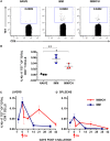
Immunization with Plasmodium berghei (Pb) radiation-attenuated sporozoite (RAS) and challenge with Pb sporozoites (spz) induced expansion of Kb-17-Tet+CD8 T cells. (A,B) B6 mice were immunized and challenged as described in Figure 1. Enriched splenic CD8 T cells from naïve, RAS immunized (IMM), and immunized/challenged (IMM/CH) mice were stained with PE-labeled H-2Kb-tetramers exchanged with the Kb-17 peptide (PBANKA_103100). (A) Representative data displayed are from single mouse and values represent the percentage of Kb-17-Tet+CD8 cells from total CD3+CD8+ population. (B) Representative results from one out of four experiments expressed as percentage of Kb-17-Tet+ CD8 T cells of total splenic CD3+CD8+ T cells and are shown individual mice with lines representing the mean and error bars indicating standard deviation. *p < 0.05; **p < 0.01; ordinary one-way ANOVA test followed by Tukey’s multiple comparisons test. (C,D) Mice immunized thrice with Pb RAS were challenged with10 K infectious Pb spz intravenously 7 days after the last boost immunization or were left unchallenged. Kinetics of (C) liver and (D) spleen Kb-17-Tet+ CD8 T cells were analyzed at the various time points, including the day of last boost immunization with Pb RAS (day −7), day of challenge (day 0), and the indicated time points thereafter. Representative results from one out of two experiments expressed as percentage of Kb-17-Tet+CD8 T cells of total liver or splenic CD8 T cells are shown as the mean of three individual mice per group, per time point.
Figure 3
Figure 3
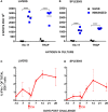
Stimulation with Kb-17 peptide induces production of IFN-γ by intrahepatic mononuclear cells (IHMC) and splenocytes from mice immunized with Plasmodium berghei (Pb) radiation-attenuated sporozoite (RAS). (A,B) IHMC and splenocytes, isolated from naïve and Pb RAS-immunized mice according to the timeline shown in Figure 1, were stimulated with Kb-17 peptide in vitro in anti-IFN-γ coated ELISPOT plates for 42 h. Unstimulated cells served as a negative control; cells stimulated with PbTRAP130 peptide served as a positive control. Representative results from two combined experiments are expressed as the net number of IFN-γ+ spots per 106 cells following subtraction of spots from unstimulated control wells. ****p < 0.0001; two-way ANOVA test followed by Sidak’s multiple comparisons test. (C,D) B6 mice were immunized by intravenously injection with 75 K Pb RAS followed by two boosts with 20 K RAS every 2 weeks and challenged with 10 K Pb sporozoites 2 weeks after the last boost. Liver and spleen cells were intracellularly stained for IFN-γ at the indicated time points. Day −14 represents the day of the third immunization, day −7 represents 7 days after the third RAS immunization, and day 0 is the day of challenge. Results are expressed as percentage of IFN-γ+ cells of total CD3+CD8+ T cells in the liver or the spleen. Results are representative of one out of two experiments and shown as the mean of three individual mice per group/per time point. Arrows indicate the day of challenge.
Figure 4
Figure 4
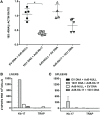
Immunization with adenovirus serotype 5 (Ad5)-Kb-17 minigene vector reduces liver stage parasite burden (LPB) after Plasmodium berghei (Pb) sporozoites (spz) challenge and induces CD8 IFN-γ responses in the spleens and livers. (A) B6 mice were primed intramuscularly with 100 µg PBANKA_103100 plasmid DNA and boosted intravenously (i.v.) with 2 × 109 infectious units Ad5-Kb-17 vector 6 weeks after the prime (or vice versa) and challenged with 10 K Pb spz i.v. Reduction of LPB was determined by qPCR in the livers 40 h after the challenge and expressed as ratio of Pb 18S rRNA to mouse β-actin. Immunization with empty plasmid DNA [empty vector (EV) DNA] and empty Ad5 (Ad5-null) served as a negative control. Results representing one out of two experiments are presented. *p < 0.01; Kruskal–Wallis test followed by Dunn’s multiple comparisons test. (B,C) Intrahepatic mononuclear cells and splenocytes were harvested 7 days after the boost immunization and stimulated with Kb-17 peptide in vitro in ELISPOT plates for 42 h. Non-stimulated cells and cells stimulated with PbTRAP130 peptide served as a negative control. Representative results of one out of three experiments are shown for pooled livers from three mice (B) or as the mean of three individual mice per group (C) and expressed as the net number of IFN-γ+ spots per million cells after subtraction of unstimulated samples.
Figure 5
Figure 5
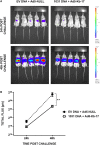
Immunization with DNA and adenovirus serotype 5 (Ad5)-Kb-17 induces reduction in liver stage parasite burden determined by in vivo imaging system. Albino-B6 mice were immunized by heterologous prime-boost with DNA and Ad5-Kb-17 at a 6-week interval as described in the legend for Figure 4 and challenged with 10 K Pb-luc sporozoites i.v. (A) Representative in vivo images of luminescence in the livers are shown at 24 and 48 h postchallenge. (B) Luminescence signal intensity in the livers at 24 and 48 h postchallenge. Representative results from one out of two experiments are shown as the mean of five individual mice per group. **p < 0.01; two-way ANOVA test followed by Sidak’s multiple comparisons test.

References

    1. WHO. World Malaria Report 2016. World Health Organization; (2016).
    1. RTS,S Clinical Trials Partnership. Agnandji ST, Lell B, Fernandes JF, Abossolo BP, Methogo BG, et al. A phase 3 trial of RTS,S/AS01 malaria vaccine in African infants. N Engl J Med (2012) 367(24):2284–95.10.1056/NEJMoa1208394 - DOI - PMC - PubMed
    1. Olotu A, Fegan G, Wambua J, Nyangweso G, Awuondo KO, Leach A, et al. Four-year efficacy of RTS,S/AS01E and its interaction with malaria exposure. N Engl J Med (2013) 368(12):1111–20.10.1056/NEJMoa1207564 - DOI - PMC - PubMed
    1. RTS,S Clinical Trials Partnership. Efficacy and safety of the RTS,S/AS01 malaria vaccine during 18 months after vaccination: a phase 3 randomized, controlled trial in children and young infants at 11 African sites. PLoS Med (2014) 11(7):e1001685.10.1371/journal.pmed.1001685 - DOI - PMC - PubMed
    1. Reece WH, Pinder M, Gothard PK, Milligan P, Bojang K, Doherty T, et al. A CD4(+) T-cell immune response to a conserved epitope in the circumsporozoite protein correlates with protection from natural Plasmodium falciparum infection and disease. Nat Med (2004) 10(4):406–10.10.1038/nm1009 - DOI - PubMed

Publication types