. 2018 Feb;15(2):1241-1246.
doi: 10.3892/etm.2017.5542. Epub 2017 Nov 22.

Assessment of dermal papillary and microvascular parameters in psoriasis vulgaris using in vivo reflectance confocal microscopy

Affiliations

Assessment of dermal papillary and microvascular parameters in psoriasis vulgaris using in vivo reflectance confocal microscopy

Alexandra Batani et al. Exp Ther Med. 2018 Feb.

Abstract

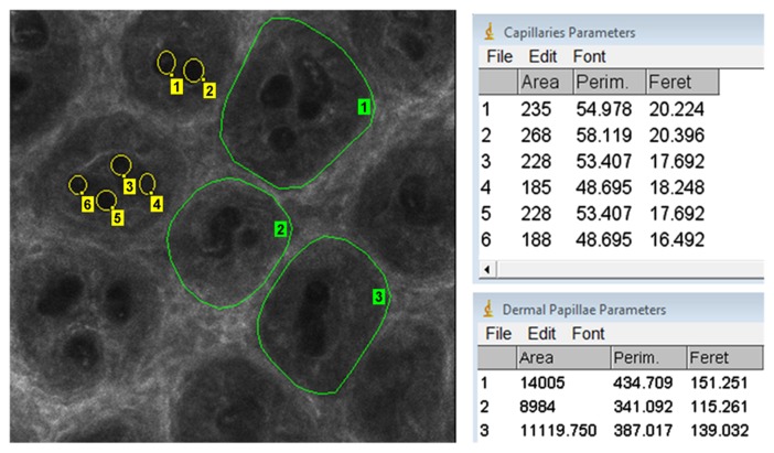
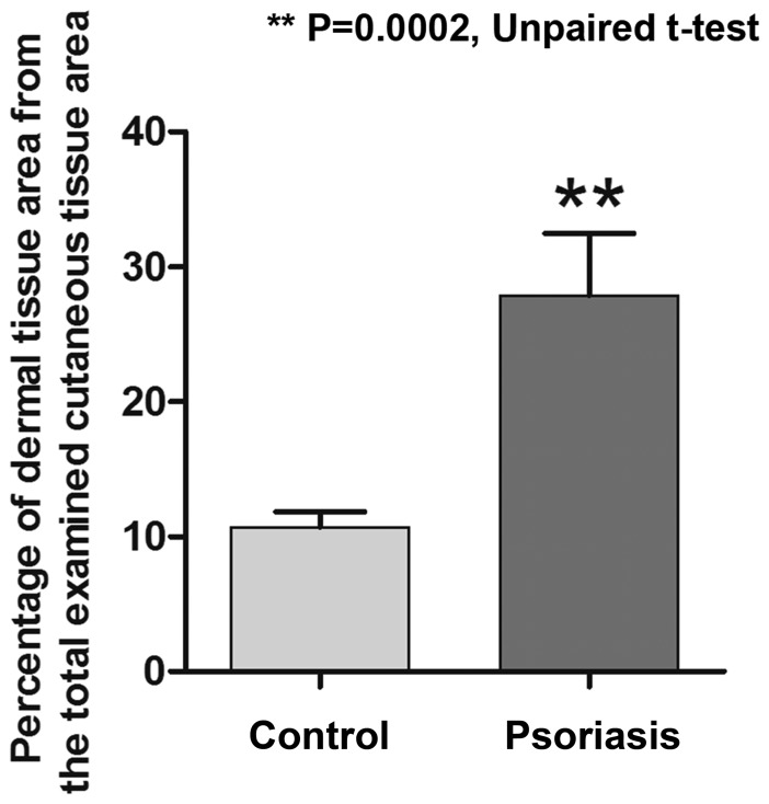
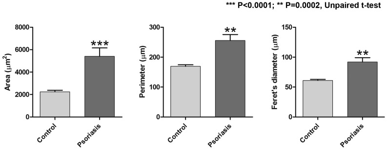
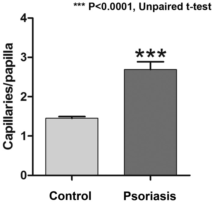
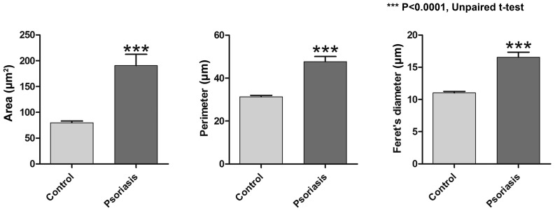
In vivo reflectance confocal microscopy (RCM) is a modern, non-invasive imaging technique, which allows for real-time examination of the upper layers of the skin at a resolution similar to that of classic microscopy. In addition, it has the advantage of real-time evaluation of blood flow and dynamic monitoring of cutaneous changes while preserving tissue integrity. The present study reported on the in vivo RCM technique as an objective method for the noninvasive assessment of psoriasis vulgaris that is potentially applicable in clinical studies and in monitoring the evolution of lesions under treatment. In psoriasis lesions, RCM virtual horizontal sections at the level of the dermo-epidermal junction featured numerous and prominent dermal papillae that were not surrounded by bright rings of basal cells. Micromorphological examination of the lesions using this technique revealed that mean values of the section area, the perimeter and the Feret's diameter of the dermal papillae were significantly higher in psoriatic lesions than in normal skin. An increased number of capillary vessels per lesional dermal papilla as compared to healthy skin was observed. Furthermore, micromorphological parameters of dermal capillaries were increased in psoriatic skin. These observations point to the utility of in vivo RCM as a promising technique for the non-invasive diagnosis of psoriasis vulgaris, for monitoring the evolution of lesions at a micromorphological level under various treatments and for gaining a better understanding of the pathophysiological processes that occur in the evolution of this disease.

Keywords: psoriasis vulgaris; reflectance confocal microscopy.


Figures

Figure 1.
Figure 1.
Micromorphological parameters of dermal papillae and dermal capillaries. The parameters of the dermal papillae (green contour lines) and of capillary vessels (yellow circles) were measured using Image J 1.45 analysis software. The parameters investigated were area, Perim and Feret. Perim, perimeter; Feret, Feret's diameter.
Figure 2.
Figure 2.
Difference between the percentage of dermal tissue from the total examined cutaneous tissue at the level of the dermo-epidermal junction. Due to the papillomatosis present in the lesional skin, a larger area of dermal tissue was also identified on the reflectance confocal microscopy images. Values are expressed as the mean ± standard deviation. **P=0.0002.
Figure 3.
Figure 3.
Comparison of parameters of dermal papillae (area, perimeter, Feret's diameter) between the two study groups. All three parameters were significantly increased in the skin affected by psoriasis, as compared with those in healthy skin. Values are expressed as the mean ± standard deviation. ***P<0.0001, **P=0.0002.
Figure 4.
Figure 4.
RCM images from psoriasis lesions and healthy skin. (A) RCM mosaic of 1.5×1.5 mm from the skin of a healthy volunteer; dermal papillae are visible in the dermo-epidermal junction as round-oval, dark areas with homogeneous structure, surrounded by bright rings of basal cells. (B) Magnified window from (A), in which capillaries are visible as round black structures (yellow arrowheads) in the center of dermal papillae (yellow asterisks). (C) RCM mosaic of 1.5×1.5 mm at the dermo-epidermal junction from the skin of a psoriasis patient. The image reveals increased size of the dermal papillae, which are separated by a thin epithelial septum, while papillary rings are absent. (D) Magnified window from C exhibiting enlarged papillae and basal cells (yellow asterisks) filled with prominent, round and dark structures that are indicative of dilated vessels (yellow arrows), while the bright rings around them in B are absent (scale bar, 300 or 100 µm). RCM, reflectance confocal microscopy.
Figure 5.
Figure 5.
Difference between skin affected by psoriasis and healthy skin regarding the mean number of capillary sections in each dermal papillae. Values are expressed as the mean ± standard deviation. ***P<0.0001.
Figure 6.
Figure 6.
Comparison of parameters of dermal capillaries (area, perimeter, Feret's diameter) between the control group and the psoriasis group. In the lesional skin, all three parameters had significantly higher values compared with those of healthy skin. Values are expressed as the mean ± standard deviation. ***P<0.0001.

Similar articles

Cited by

References

    1. Naldi L. Epidemiology of psoriasis. Curr Drug Targets Inflamm Allergy. 2004;3:121–128. doi: 10.2174/1568010043343958. - DOI - PubMed
    1. Parisi R, Symmons DP, Griffiths CE, Ashcroft DM, Identification and Management of Psoriasis and Associated ComorbidiTy (IMPACT) project team Global epidemiology of psoriasis: A systematic review of incidence and prevalence. J Invest Dermatol. 2013;133:377–385. doi: 10.1038/jid.2012.339. - DOI - PubMed
    1. Caruntu C, Grigore C, Caruntu A, Diaconeasa A, Boda D. The role of stress in skin diseases. Med Interna (Internal Med) 2011;8:73–84.
    1. Villarreal-Martínez A, Gallardo-Blanco H, Cerda-Flores R, Torres-Muñoz I, Gómez-Flores M, Salas-Alanís J, Ocampo-Candiani J, Martínez-Garza L. Candidate gene polymorphisms and risk of psoriasis: A pilot study. Exp Ther Med. 2016;11:1217–1222. doi: 10.3892/etm.2016.3066. - DOI - PMC - PubMed
    1. Chen W, Xie K, Liu X, Chen H. Identification of key pathways and genes in psoriasis via gene microarray analysis. Mol Med Rep. 2016;13:2327–2337. - PubMed