

Function and mechanism of microRNA-210 in acute cerebral infarction
- PMID: 29434712
- PMCID: PMC5774459
- DOI: 10.3892/etm.2017.5577
Function and mechanism of microRNA-210 in acute cerebral infarction
Abstract
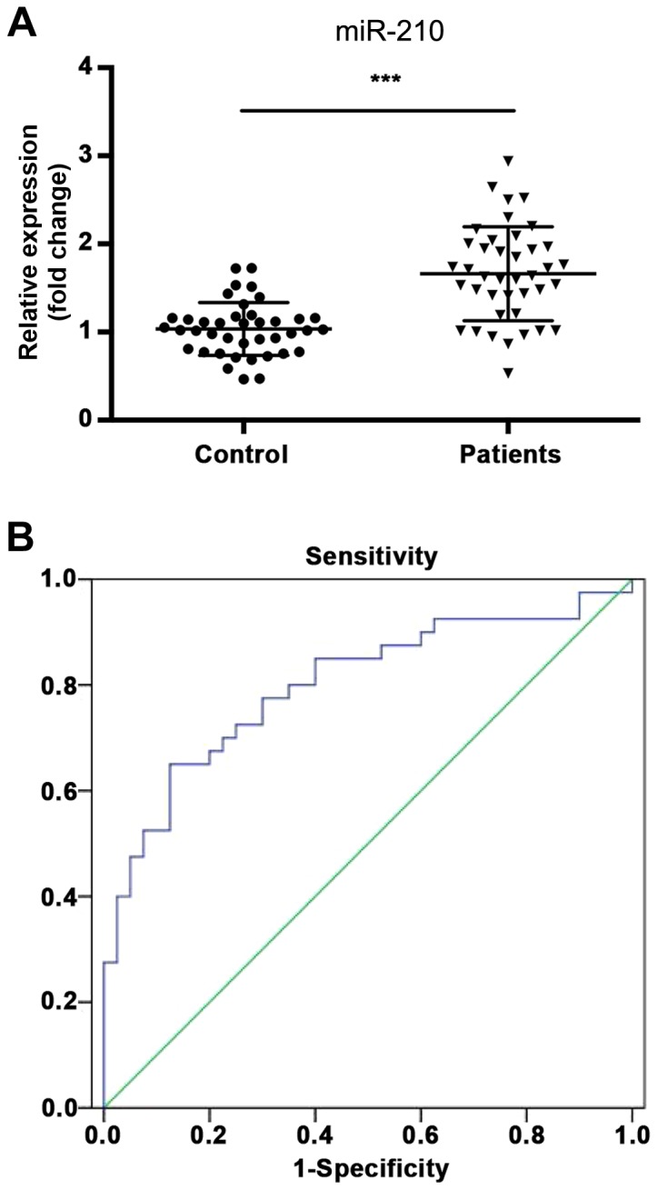
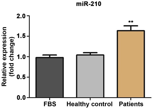
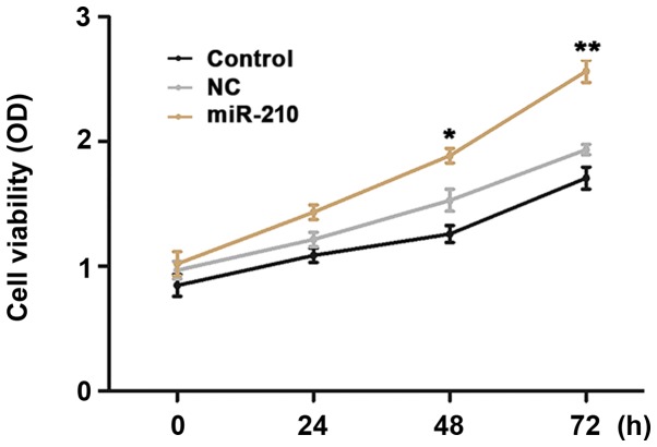
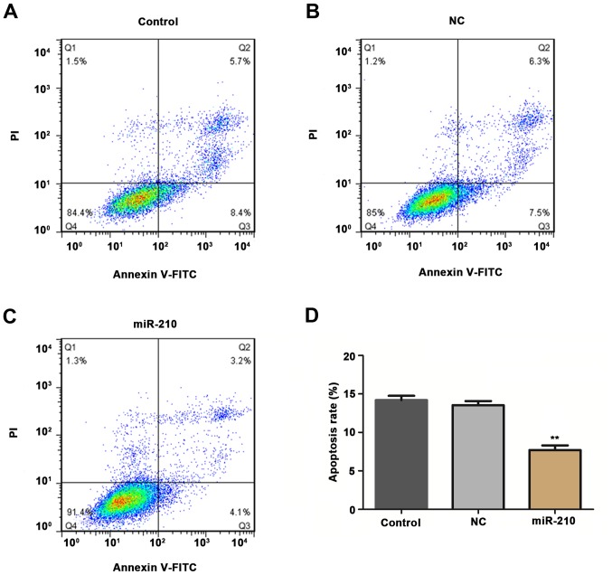
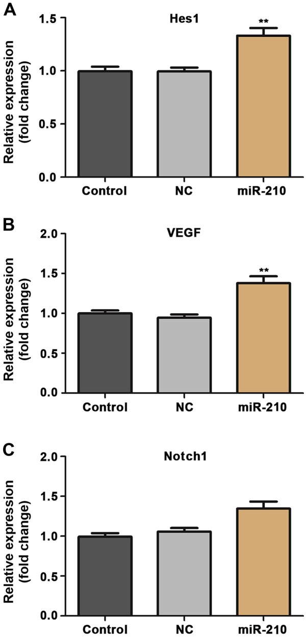
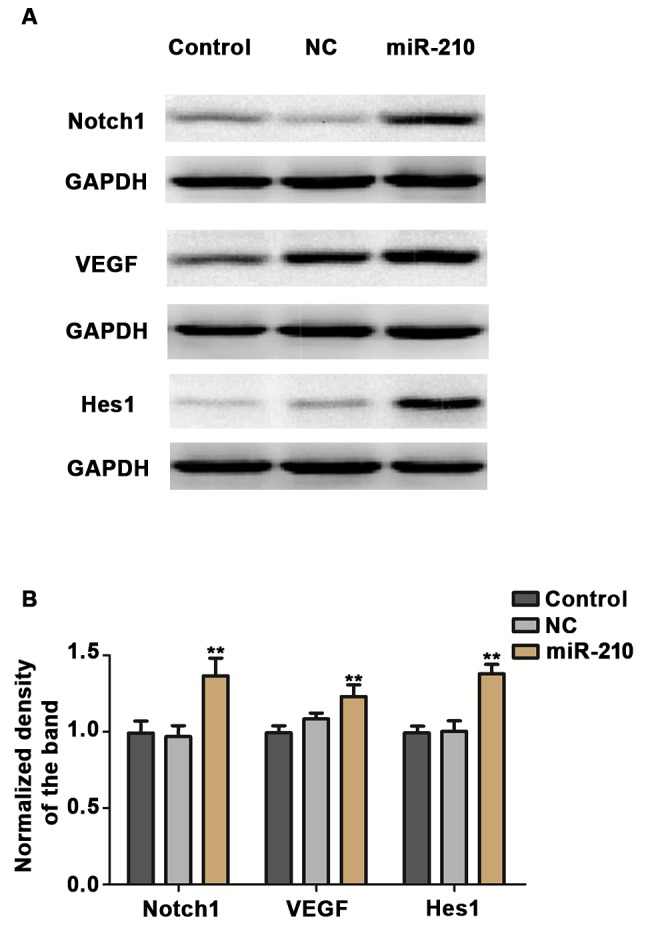
Acute cerebral infarction (ACI) is a common cerebrovascular disease. Previous studies have indicated that microRNAs (miRs) are aberrantly expressed in patients with ACI. However, the functions of miRs in the pathogenesis of ACI still require further investigation. The aim of the present study was to investigate the function of miR-210 in ACI and its associated mechanism. The expression of miR-210 in the serum of 40 patients with ACI and 40 normal controls was examined using reverse transcription-quantitative polymerase chain reaction (RT-qPCR). Then, human umbilical vein endothelial cells (HUVECs) were treated with serum from patients with ACI or healthy volunteers, and a CCK-8 assay was performed to examine cell proliferation. Next, cells were stained with PI/Annexin V, and the apoptosis rate was examined using flow cytometry. Furthermore, cells were harvested and lysed, and RT-qPCR and western blotting assays were performed to compare the expression of vascular endothelial growth factor (VEGF), Notch1 and Hes1 in different groups. It was observed that the expression of miR-210 was significantly increased in the serum of patients with ACI compared with normal controls (P<0.01), and receiver operating characteristic curve analysis indicated that the area under the curve for miR-210 was 0.799 (95% confidence interval, 0.700-0.899), the optimum cut-off point was 1.397, and the sensitivity and specificity at the cut-off point were 62.5 and 87.5%, respectively. Furthermore, serum from patients with ACI induced a significant increase in proliferation (P<0.05 at 48 h, P<0.01 at 72 h) and a significant decrease in the apoptosis rate of HUVECs (P<0.01). In addition, serum from patients with ACI significantly increased the expression of VEGF, Notch1 and Hes1 at the mRNA and protein level (all P<0.01 with the exception of Notch1 mRNA expression, P>0.05). In conclusion, these results demonstrate that miR-210 is upregulated in the serum of patients with ACI, and miR-210 may be involved in the pathogenesis of ACI through regulating the proliferation and apoptosis of endothelial cells.
Keywords: acute cerebral infarction; apoptosis; human umbilical vein endothelial cells; micoRNA-210; proliferation.
Figures
Similar articles
-
Abnormal expression of miRNA-122 in cerebral infarction and related mechanism of regulating vascular endothelial cell proliferation and apoptosis by targeting CCNG1.Clinics (Sao Paulo). 2023 Apr 27;78:100199. doi: 10.1016/j.clinsp.2023.100199. eCollection 2023. Clinics (Sao Paulo). 2023. PMID: 37119591 Free PMC article.
-
[Expression and its significance of microRNA-210 in serum in acute cerebral infarction].Zhonghua Wei Zhong Bing Ji Jiu Yi Xue. 2014 Dec;26(12):910-3. doi: 10.3760/cma.j.issn.2095-4352.2014.12.013. Zhonghua Wei Zhong Bing Ji Jiu Yi Xue. 2014. PMID: 25476086 Chinese.
-
Plasma level of miR-99b may serve as potential diagnostic and short-term prognostic markers in patients with acute cerebral infarction.J Clin Lab Anal. 2020 Mar;34(3):e23093. doi: 10.1002/jcla.23093. Epub 2020 Jan 22. J Clin Lab Anal. 2020. PMID: 31967688 Free PMC article.
-
[The expression of serummiR-151a-3p in patients with acute cerebral infarction and its correlation with pro-inflammatory factors].Zhonghua Wei Zhong Bing Ji Jiu Yi Xue. 2016 Mar;28(3):272-6. Zhonghua Wei Zhong Bing Ji Jiu Yi Xue. 2016. PMID: 29917348 Chinese.
-
Up-regulation of microRNA-503 by high glucose reduces the migration and proliferation but promotes the apoptosis of human umbilical vein endothelial cells by inhibiting the expression of insulin-like growth factor-1 receptor.Eur Rev Med Pharmacol Sci. 2018 Jun;22(11):3515-3523. doi: 10.26355/eurrev_201806_15178. Eur Rev Med Pharmacol Sci. 2018. PMID: 29917206
Cited by
-
MicroRNA-210 Downregulates TET2 (Ten-Eleven Translocation Methylcytosine Dioxygenase 2) and Contributes to Neuroinflammation in Ischemic Stroke of Adult Mice.Stroke. 2023 Mar;54(3):857-867. doi: 10.1161/STROKEAHA.122.041651. Epub 2023 Feb 3. Stroke. 2023. PMID: 36734233 Free PMC article.
-
Characteristics of Fall-Related Fractures in Older Adults with Cerebrovascular Disease: A Cross-Sectional Study.Clin Interv Aging. 2021 Jul 13;16:1337-1346. doi: 10.2147/CIA.S316739. eCollection 2021. Clin Interv Aging. 2021. PMID: 34285478 Free PMC article.
-
A systematic review of the research progress of non-coding RNA in neuroinflammation and immune regulation in cerebral infarction/ischemia-reperfusion injury.Front Immunol. 2022 Oct 7;13:930171. doi: 10.3389/fimmu.2022.930171. eCollection 2022. Front Immunol. 2022. PMID: 36275741 Free PMC article.
-
Considering Context-Specific microRNAs in Ischemic Stroke with Three "W": Where, When, and What.Mol Neurobiol. 2024 Oct;61(10):7335-7353. doi: 10.1007/s12035-024-04051-5. Epub 2024 Feb 21. Mol Neurobiol. 2024. PMID: 38381296 Review.
-
Expression and significance of S-100β, CysC and NF-κB in patients with acute cerebral infarction.Exp Ther Med. 2021 Feb;21(2):149. doi: 10.3892/etm.2020.9580. Epub 2020 Dec 16. Exp Ther Med. 2021. PMID: 33456516 Free PMC article.
References
LinkOut - more resources
Full Text Sources
Other Literature Sources
Research Materials
Miscellaneous