. 2018 Feb;15(2):2079-2084.
doi: 10.3892/ol.2017.7546. Epub 2017 Dec 7.

Extranodal presentation of Hodgkin's lymphoma of the sternum: A case report and review of the literature

Affiliations

Extranodal presentation of Hodgkin's lymphoma of the sternum: A case report and review of the literature

Yan Li et al. Oncol Lett. 2018 Feb.

Abstract

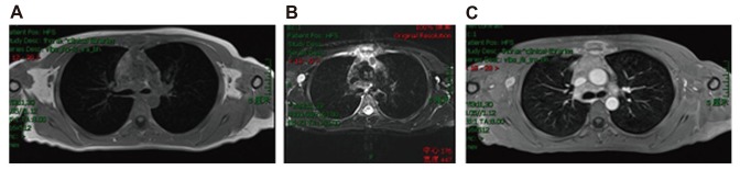
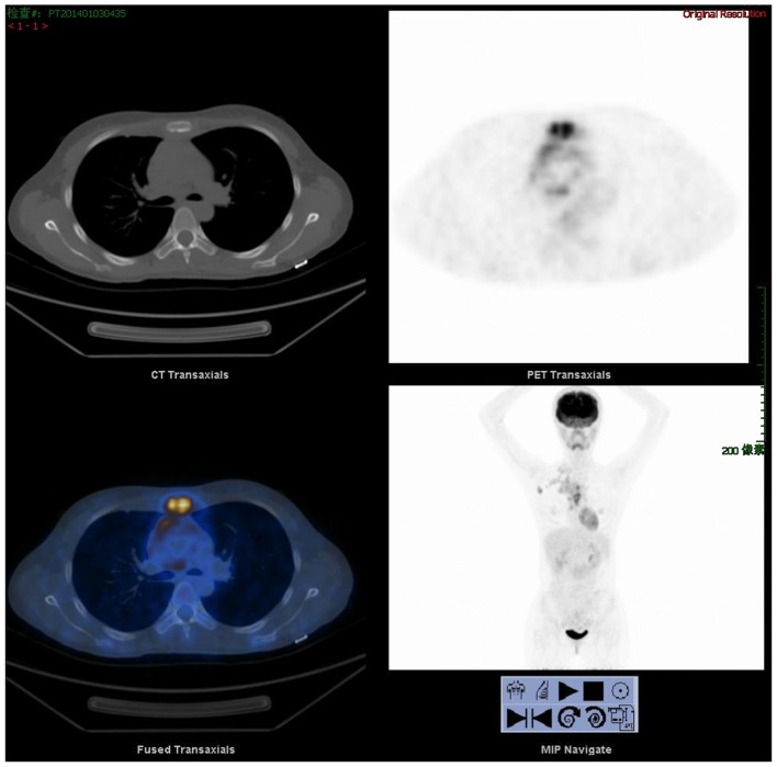
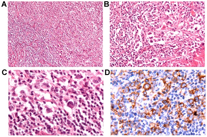
Hodgkin's lymphoma (HL) is typically a systemic disease with involvement of the cervical, supraclavicular and mediastinal lymph nodes. An extra-nodal presentation of HL is unusual and seldom encountered in the clinic. The most common sites of presentation for extra-nodal extension are the spleen, liver, lungs, bones and marrow. The bones that are frequently involved are the vertebrae, pelvis, ribs and femur. Involvement of the sternum has occasionally been reported. The current study presents an unusual case on the extra-nodal presentation of HL of the sternum arising in a 25-year-old woman, and reviews the relevant literature with particular emphasis on treatment. The extra-nodal infiltration of HL, and the clinical stage and prognosis of the case are also discussed.

Keywords: Hodgkin's lymphoma; diagnose; osseous involvement; treatment.


Figures

Figure 1.
Figure 1.
Axial magnetic resonance imaging of the lesions of the sternum. (A) T1-weighted image. (B) Enhanced scan of T1-weighted image demonstrating the discontinuous cortex of the posterior midsternum and the soft-tissue mass around. (C) T2-weighted image.
Figure 2.
Figure 2.
Positron emission tomography/computed tomography at the time of presentation demonstrating increased fluorodeoxyglucose uptake in the sternum.
Figure 3.
Figure 3.
(A) Background mixture of eosinophils, plasmacytes, lymphocytes and atypical cells. (B) Typical R-S cells are apparent. (C) Occasional lacunar cells are also apparent. (D) The large cells are positive for cluster of differentiation 30 found on the surface of R-S cells. R-S, Reed-Sternberg.

Similar articles

Cited by

References

    1. Guermazi A, Brice P, de Kerviler EE, Fermé C, Hennequin C, Meignin V, Frija J. Extranodal Hodgkin disease: Spectrum of disease. Radiographics. 2001;21:161–179. doi: 10.1148/radiographics.21.1.g01ja02161. - DOI - PubMed
    1. Zucca E. Extranodal lymphoma: A reappraisal. Ann Oncol. 2008;19(Suppl 4):iv77–iv80. doi: 10.1093/annonc/mdn204. - DOI - PubMed
    1. Chan KW, Rosen G, Miller DR, Tan CT. Hodgkin's diseases in adolescents presenting as a primary bone lesion. A report of four cases and review of literature. Am J Pediatr Hematol Oncol. 1982;4:11–17. - PubMed
    1. Biswas A, Puri T, Goyal S, Haresh KP, Gupta R, Julka PK, Rath GK. Osseous Hodgkin's lymphoma-review of literature and report of an unusual case presenting as a large ulcerofungating sternal mass. Bone. 2008;43:636–640. doi: 10.1016/j.bone.2008.05.020. - DOI - PubMed
    1. The International Non-Hodgkin's Lymphoma Prognostic Factors Project: A predictive model for aggressive non-Hodgkin's lymphoma. N Engl J Med. 1993;329:987–994. doi: 10.1056/NEJM199309303291402. - DOI - PubMed