

Overexpression of β1 integrin contributes to polarity reversal and a poor prognosis of breast invasive micropapillary carcinoma
- PMID: 29435106
- PMCID: PMC5796977
- DOI: 10.18632/oncotarget.22774
Overexpression of β1 integrin contributes to polarity reversal and a poor prognosis of breast invasive micropapillary carcinoma
Abstract
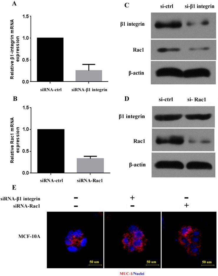
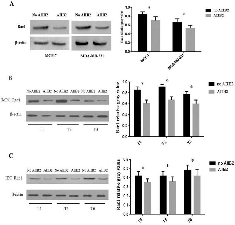
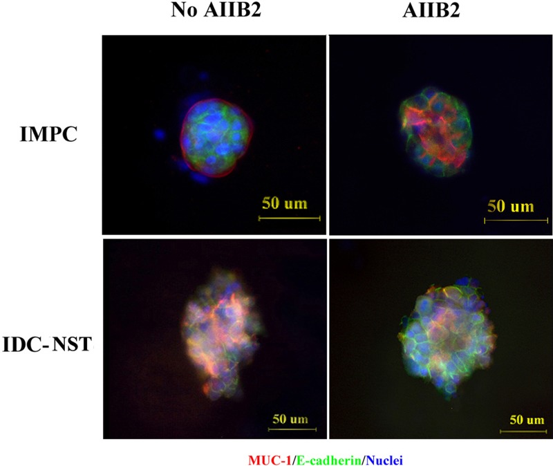
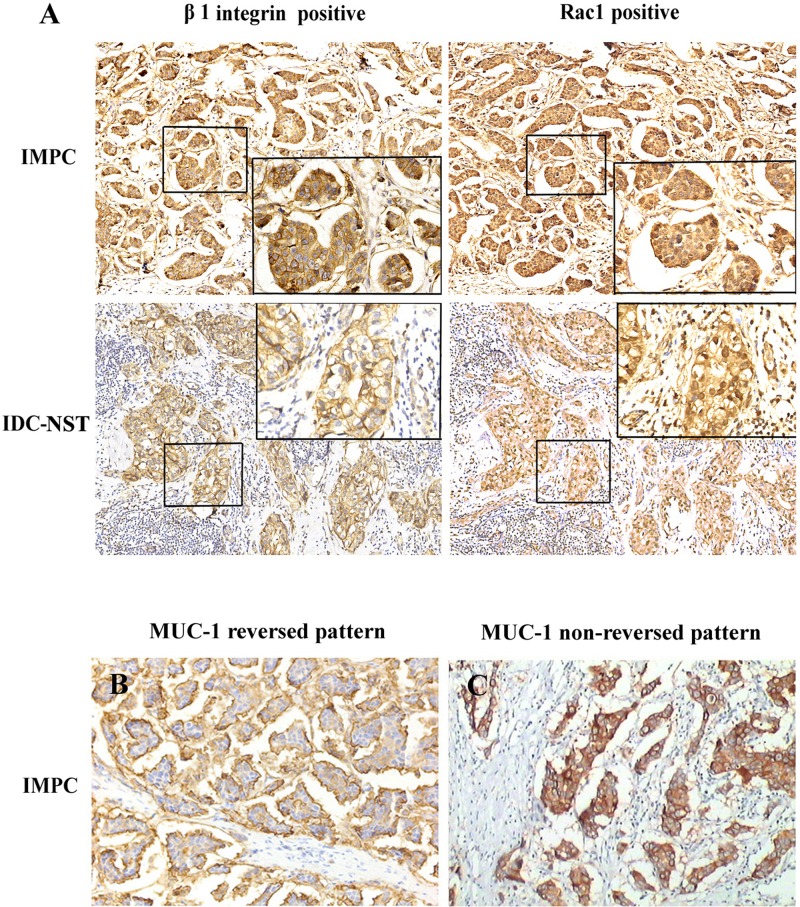
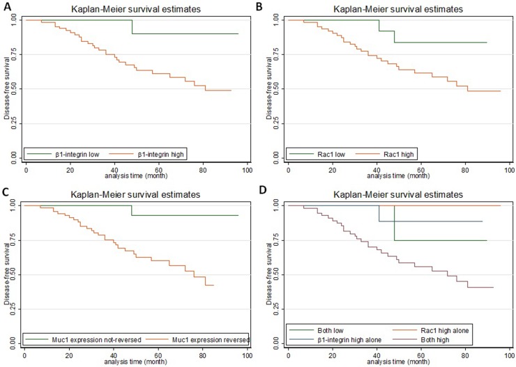
Invasive micropapillary carcinoma (IMPC) of the breast is a highly aggressive breast cancer. Polarity reversal exemplified by cluster growth is hypothesized to contribute to the invasiveness and metastasis of IMPC. In this study, we demonstrate that levels of β1 integrin and Rac1 expression were greater in breast IMPC than in invasive breast carcinoma of no specific type and paraneoplastic benign breast tissue. We show that silencing β1 integrin expression using the β1 integrin inhibitor AIIB2 partially restored polarity in IMPC primary cell clusters and downregulated Rac1. Thus, overexpression of β1 integrin upregulates Rac1. Univariate analysis showed that overexpression of β1 integrin and Rac1 was associated with breast cancer cell polarity reversal, lymph node metastasis, and poor disease-free survival in IMPC patients. Multivariate analysis revealed that polarity reversal was an independent predictor of poor disease-free survival. These findings indicate that overexpression of β1 integrin and the resultant upregulation of Rac1 contribute to polarity reversal and metastasis of breast IMPC, and that β1 integrin and Rac1 could be potential prognostic biomarkers and targets for treatment of breast IMPC.
Keywords: breast; invasive micropapillary carcinoma; metastasis; polarity reversal; β1 integrin.
Conflict of interest statement
CONFLICTS OF INTEREST None.
Figures
References
-
- Amin MB, Ro JY, el-Sharkawy T, Lee KM, Troncoso P, Silva EG, Ordóñez NG, Ayala AG. Micropapillary variant of transitional cell carcinoma of the urinary bladder: histologic pattern resembling ovarian papillary serous carcinoma. Am J Surg Pathol. 1994;18:1224–1232. - PubMed
-
- Sakamoto K, Watanabe M, De La Cruz C, Honda H, Ise H, Mitsui K, Namiki K, Mikami Y, Moriya T, Sasano H. Primary invasive micropapillary carcinoma of the colon. Histopathology. 2005;47:479–484. - PubMed
-
- Kuroda N, Hamaguchi N, Ohara M, Hirouchi T, Miyzaki E, Mizuno K. Intracytoplasmic lumina in invasive micropapillary carcinoma of the lung. Diagn Cytopathol. 2006;34:224–226. - PubMed
-
- Fisher ER, Palekar AS, Redmond C, Barton B, Fisher B. Pathologic findings from the National Surgical Adjuvant Breast Project (protocol no. 4): VI, invasive papillary cancer. Am J Clin Pathol. 1980;73:313–322. - PubMed
-
- Tavessoli FA, Devilee P. WHO Classification of Tumours: Pathology and Genetics: Tumours of the Breast and Female Genital Organs. Lyon, France: IARC Press; 2003. p. 10.
LinkOut - more resources
Full Text Sources
Other Literature Sources
Research Materials
