. 2017 Dec 20;9(4):4707-4721.
doi: 10.18632/oncotarget.23523. eCollection 2018 Jan 12.

Epigenetic silencing of SMOC1 in traditional serrated adenoma and colorectal cancer

Affiliations

Epigenetic silencing of SMOC1 in traditional serrated adenoma and colorectal cancer

Hironori Aoki et al. Oncotarget. .

Abstract

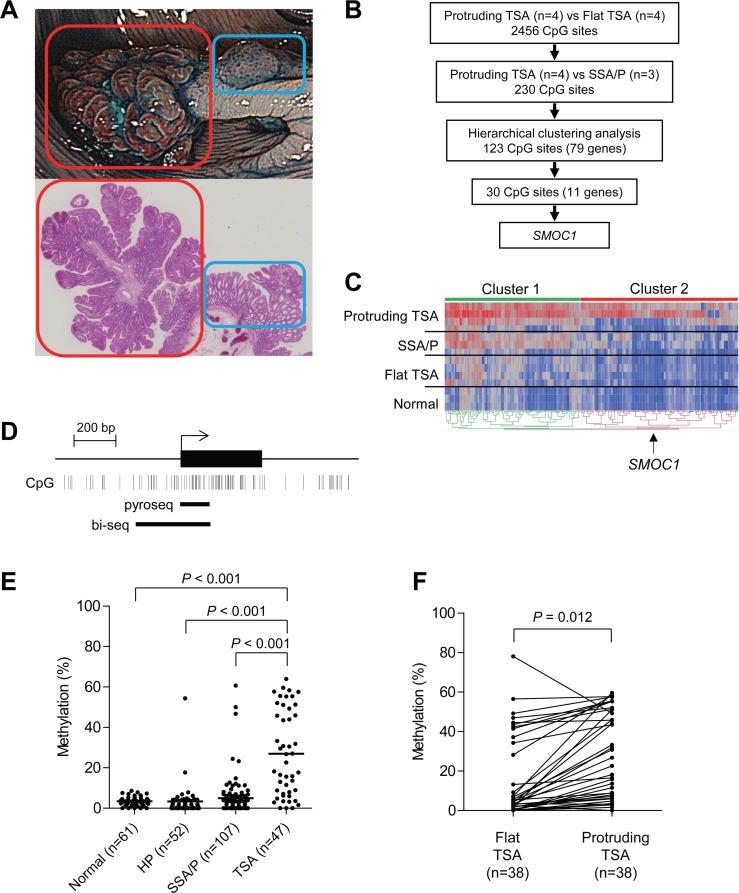
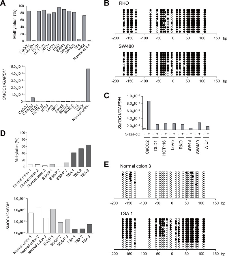
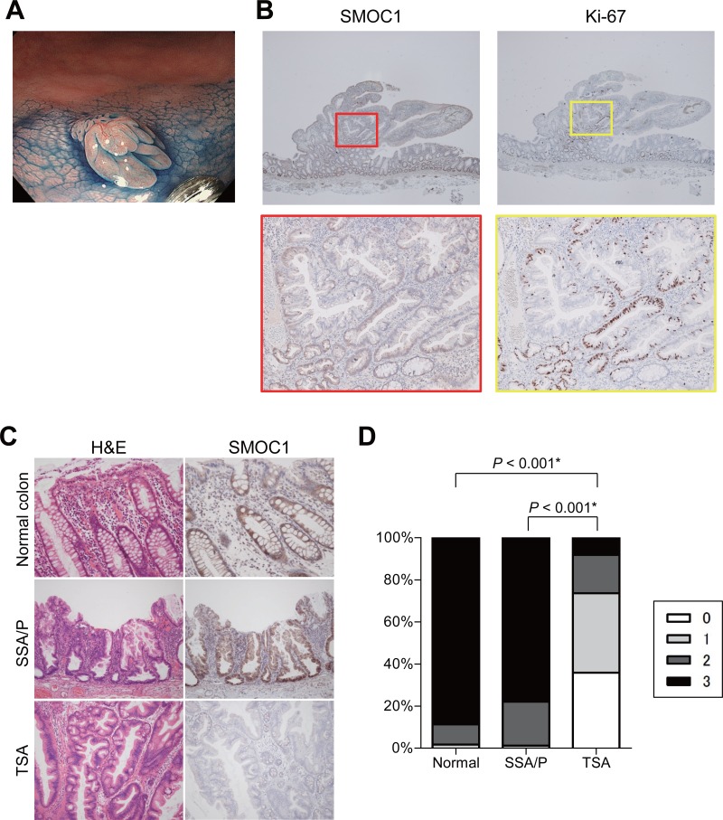
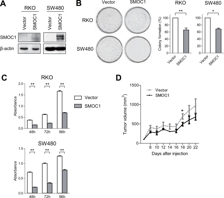
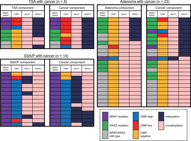
Colorectal sessile serrated adenoma/polyps (SSA/Ps) are well-known precursors of colorectal cancer (CRC) characterized by BRAF mutation and microsatellite instability. By contrast, the molecular characteristics of traditional serrated adenoma (TSAs) are not fully understood. We analyzed genome-wide DNA methylation in TSAs having both protruding and flat components. We identified 11 genes, including SMOC1, methylation of which progressively increased during the development of TSAs. SMOC1 was prevalently methylated in TSAs, but was rarely methylated in SSA/Ps (p < 0.001). RT-PCR and immunohistochemistry revealed that SMOC1 was expressed in normal colon and SSA/Ps, but its expression was decreased in TSAs. Ectopic expression of SMOC1 suppressed proliferation, colony formation and in vivo tumor formation by CRC cells. Analysis of colorectal lesions (n = 847) revealed that SMOC1 is frequently methylated in TSAs, high-grade adenomas and CRCs. Among these, SMOC1 methylation was strongly associated with KRAS mutation and CpG island methylator phenotype (CIMP)-low. These results demonstrate that epigenetic silencing of SMOC1 is associated with TSA development but is rarely observed in SSA/Ps. SMOC1 expression could thus be a diagnostic marker of serrated lesions, and SMOC1 methylation could play a role in neoplastic pathways in TSAs and conventional adenomas.

Keywords: CIMP; DNA methylation; SMOC1; colorectal cancer; traditional serrated adenoma.


Conflict of interest statement

CONFLICTS OF INTEREST All authors declare no conflict of interest.

Figures

Figure 1
Figure 1. Identification of DNA methylation associated with the development of TSAs
(A) Representative example of TSA. An endoscopic view is shown on the top and a histological view is below. Red boxes indicate a protruding component and blue boxes indicate a flat component. (B) Workflow to identify DNA methylation in TSAs. (C) Heatmap of the selected 230 CpG sites within the protruding components of TSAs (n = 4), SSA/Ps (n = 3), flat components of TSAs (n = 4) and normal colonic tissue (n = 3). Clusters 1 and 2 include 107 and 123 CpG sites, respectively. (D) Diagram of the promoter region of SMOC1. The transcription start site and exon 1 are shown on the top, and the regions analyzed using bisulfite pyrosequencing and bisulfite sequencing are shown below. (E) Summarized results of bisulfite pyrosequencing of SMOC1 in specimens from the indicated lesions and adjacent normal colonic tissues. (F) Results of bisulfite pyrosequencing of SMOC1 in specimens from TSAs consisting of protruding and flat components.
Figure 2
Figure 2. Analysis of SMOC1 methylation and expression in colorectal tumors
(A) Bisulfite pyrosequencing (top) and qRT-PCR (bottom) analyses of SMOC1 in the indicated CRC cell lines and normal colonic tissue. (B) Bisulfite sequencing analysis of the SMOC1 CpG island in the indicated CRC cell lines. Open and filled circles represent unmethylated and methylated CpG sites, respectively. (C) qRT-PCR analysis of SMOC1 in the indicated CRC cell lines, with or without 5-aza-dC treatment. (D) Bisulfite pyrosequencing (top) and qRT-PCR (bottom) analyses of SMOC1 in normal colonic tissues and primary serrated lesions. (E) Bisulfite sequencing analysis of SMOC1 in the representative samples in (D).
Figure 3
Figure 3. Immunohistochemical analysis of SMOC1 in serrated lesions
(A) Endoscopic view of a TSA with protruding and flat components. (B) SMOC1 (left) and Ki-67 (right) staining in the TSA specimen shown in (A). Magnified views indicated by red and yellow boxes are shown below. (C) Representative views of hematoxylin and eosin (left) and SMOC1 (right) staining in normal colonic tissue, SSA/P and TSA specimens. (D) Summarized results for SMOC1 expression levels in normal colon (n = 23), SSA/Ps (n = 12) and TSAs (n = 11). *Fisher's exact test.
Figure 4
Figure 4. Functional analysis of SMOC1 in CRC cells
(A) Western blot analysis of SMOC1 in the indicated CRC cells transfected with a SMOC1 expression vector or a control vector (Vector). (B) Colony formation assays using the indicated CRC cell lines transfected with the indicated vectors. Representative results are on the left, and relative colony formation efficiencies are on the right. Shown are means of 3 replications; error bars represent SDs. *P < 0.05, **P < 0.01. (C) Cell viability assays using the indicated CRC cell lines transfected with the indicated vectors. Shown are means of 8 replications; error bars represent SDs. **P < 0.01. (D) Tumor growth in mice injected with SW480 cells transfected with the indicated vectors. Shown are means of 5 replications; error bars represent SDs. *P < 0.05.
Figure 5
Figure 5. Changes in the molecular alterations during the progression of colorectal tumors
Summary of the molecular features in colorectal tumors consisting of premalignant and malignant components are shown.

References

    1. Fearon ER, Vogelstein B. A genetic model for colorectal tumorigenesis. Cell. 1990;61:759–767. - PubMed
    1. Leggett B, Whitehall V. Role of the serrated pathway in colorectal cancer pathogenesis. Gastroenterology. 2010;138:2088–2100. - PubMed
    1. Bosman FT, World Health Organization . WHO classification of tumours of the digestive system. Lyon:: International Agency for Research on Cancer; 2010. International Agency for Research on Cancer.
    1. Spring KJ, Zhao ZZ, Karamatic R, Walsh MD, Whitehall VL, Pike T, Simms LA, Young J, James M, Montgomery GW, Appleyard M, Hewett D, Togashi K, et al. High prevalence of sessile serrated adenomas with BRAF mutations: a prospective study of patients undergoing colonoscopy. Gastroenterology. 2006;131:1400–1407. - PubMed
    1. Kimura T, Yamamoto E, Yamano HO, Suzuki H, Kamimae S, Nojima M, Sawada T, Ashida M, Yoshikawa K, Takagi R, Kato R, Harada T, Suzuki R, et al. A novel pit pattern identifies the precursor of colorectal cancer derived from sessile serrated adenoma. Am J Gastroenterol. 2012;107:460–469. - PubMed

LinkOut - more resources