Review
. 2015 Nov 1;6(11):6059-6068.
doi: 10.1039/c5sc02546j. Epub 2015 Aug 26.

Excitonic splittings in molecular dimers: why static ab initio calculations cannot match them

Affiliations
Review

Excitonic splittings in molecular dimers: why static ab initio calculations cannot match them

Philipp Ottiger et al. Chem Sci. .

Abstract

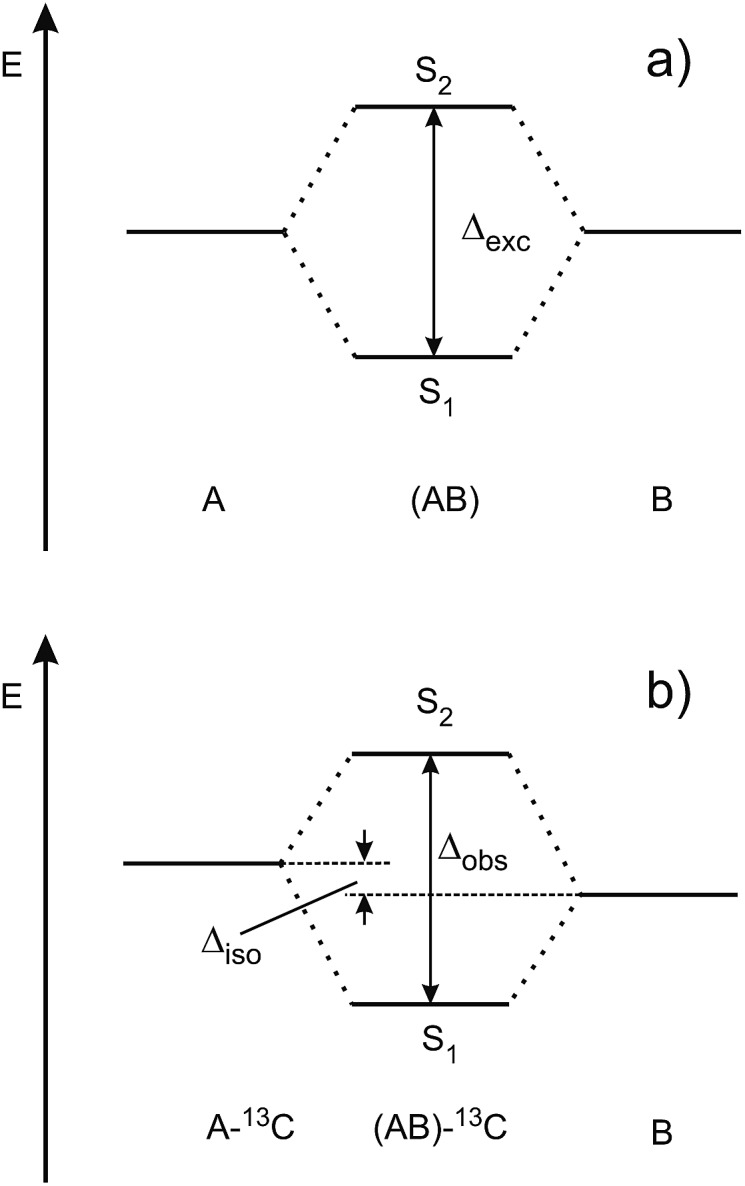
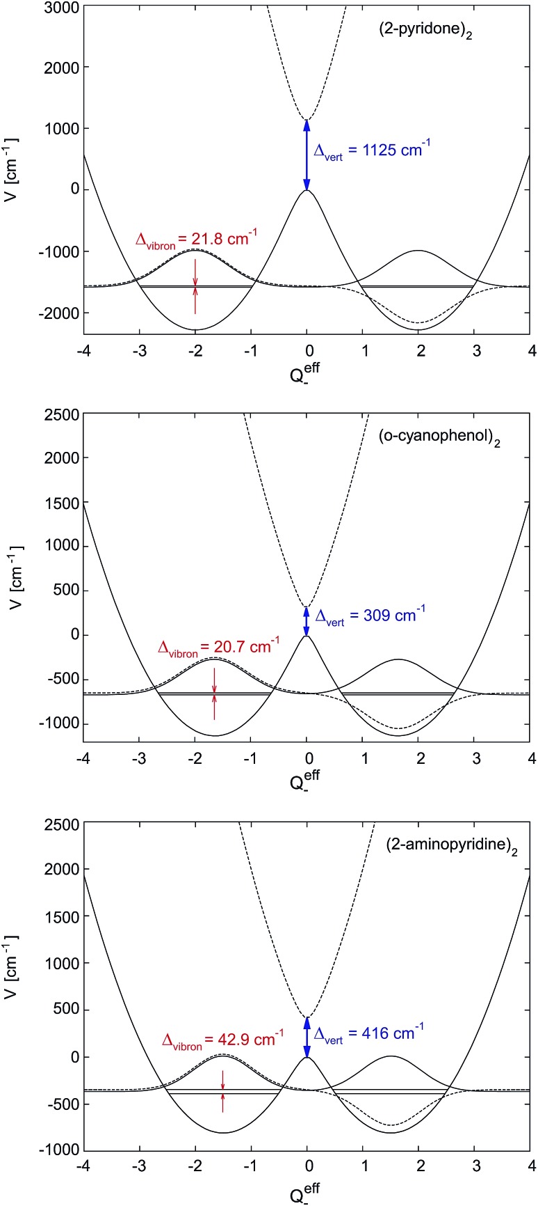
After decades of research on molecular excitons, only few molecular dimers are available on which exciton and vibronic coupling theories can be rigorously tested. In centrosymmetric H-bonded dimers consisting of identical (hetero)aromatic chromophores, the monomer electronic transition dipole moment vectors subtract or add, yielding S0 → S1 and S0 → S2 transitions that are symmetry-forbidden or -allowed, respectively. Symmetry breaking by 12C/13C or H/D isotopic substitution renders the forbidden transition weakly allowed. The excitonic coupling (Davydov splitting) can then be measured between the S0 → S1 and S0 → S2 vibrationless bands. We discuss the mass-specific excitonic spectra of five H-bonded dimers that are supersonically cooled to a few K and investigated using two-color resonant two-photon ionization spectroscopy. The excitonic splittings Δcalc predicted by ab initio methods are 5-25 times larger than the experimental excitonic splittings Δexp. The purely electronic ab initio splittings need to be reduced ("quenched"), reflecting the coupling of the electronic transition to the optically active vibrations of the monomers. The so-called quenching factors Γ < 1 can be determined from experiment (Γexp) and/or calculation (Γcalc). The vibronically quenched splittings Γ·Δcalc are found to nicely reproduce the experimental exciton splittings.


Figures

Fig. 1
Fig. 1. Ground state geometries of the H-bonded dimers (2-pyridone)2, (o-cyanophenol)2, (2-aminopyridine)2, (benzonitrile)2 and (benzoic acid)2 (CC2/aug-cc-pVTZ calculations). The monomer transition-dipole moment vectors are indicated as red double-headed arrows.
Fig. 2
Fig. 2. Schematic view of (a) the excitonic splitting in a symmetric dimer consisting of identical chromophores A and B, (b) the S1/S2 state splitting in a symmetry-broken dimer that is isotopically substituted in chromophore A.
Fig. 3
Fig. 3. Experimentally observed splittings Δobs between the S1 and S2 electronic origins of the 13C-isotopomers of the doubly H-bonded dimers in Fig. 1.
Fig. 4
Fig. 4. Two-dimensional potential energy surfaces of the S1 (red) and S2 (blue) states of a symmetric self-dimer, plotted as a function of the intramolecular vibrational coordinates QA and QB. (a) Strong coupling case, (b) weak coupling case. The calculated vibronic band patterns are shown as stickplots in (c and d), with negative band intensities (red) for Ag vibronic transitions and positive (blue) intensities for the Bu vibronic transitions.
Fig. 5
Fig. 5. Dependence of the S1/S2 intensity ratio on the excitonic splitting in H-bonded symmetric molecular dimers. The excitonic splittings were determined from the directly observable splittings of the 13C isotopomers, assuming a 12C/13C isotopic shift Δiso of 3.3 cm–1 for all dimers. The theoretical dependence of the S1/S2 intensity ratio on Δexc is indicated in red.
Fig. 6
Fig. 6. 1D-cuts of the first two excited adiabatic potential energy surfaces along the effective mode for (2-pyridone)2, (o-cyanophenol)2 and (2-aminopyridine)2. The line type of the vibronic wave functions (schematic drawing) is the same as that of the corresponding potential energy curve, its zero is chosen to match its vibronic energy. The excitation energy splitting at the ground state equilibrium geometry (Qeff– = 0) equals the electronic excitonic splitting Δvert.

References

    1. Cheng Y. C., Fleming G. Annu. Rev. Phys. Chem. 2009;60:241–262. - PubMed
    1. Novoderezhkin V. I., van Grondelle R. Phys. Chem. Chem. Phys. 2010;12:7352–7365. - PubMed
    1. Scholes G. D., Fleming G. R., Olaya-Castro A., van Grondelle R. Nat. Chem. 2011;3:763–774. - PubMed
    1. Fassioli F., Dinshaw R., Arpin P. C., Scholes G. D. J. R. Soc., Interface. 2014;11:20130901. - PMC - PubMed
    1. Strümpfer J., Schulten K. J. Chem. Phys. 2012;137:065101. - PMC - PubMed

LinkOut - more resources