Review
. 2018 Jun;23(4):599-612.
doi: 10.1007/s00775-018-1538-8. Epub 2018 Feb 12.

The unique fold and lability of the [2Fe-2S] clusters of NEET proteins mediate their key functions in health and disease

Affiliations
Review

The unique fold and lability of the [2Fe-2S] clusters of NEET proteins mediate their key functions in health and disease

Ola Karmi et al. J Biol Inorg Chem. 2018 Jun.

Abstract

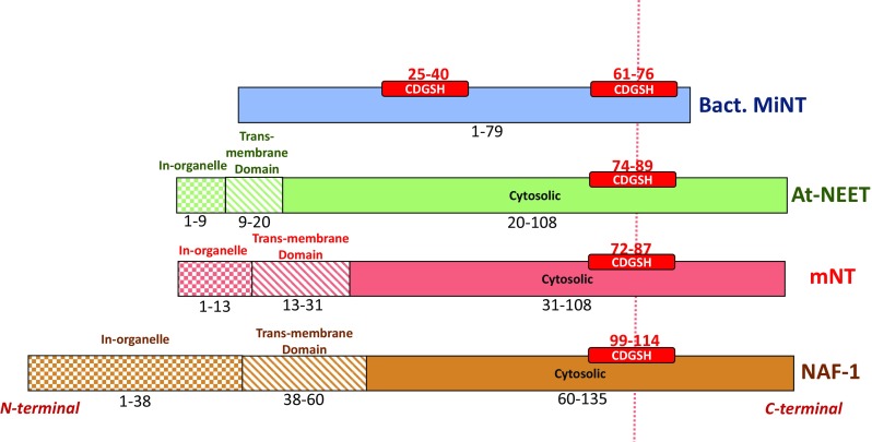
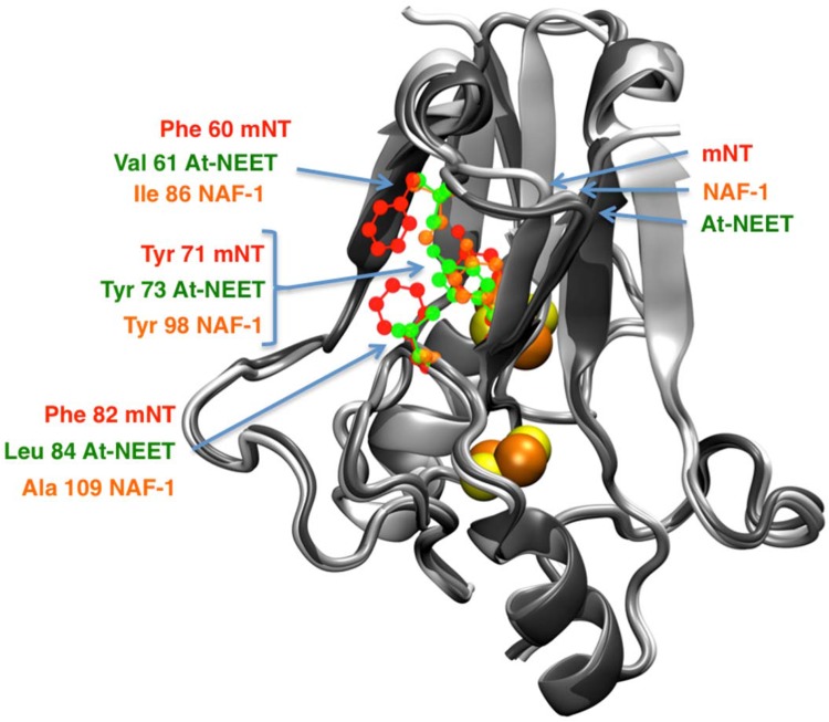
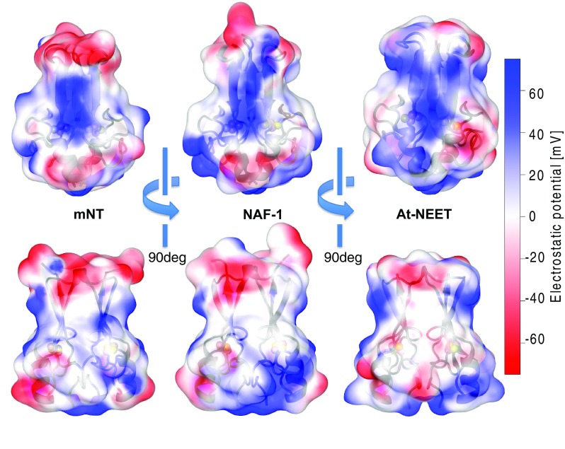
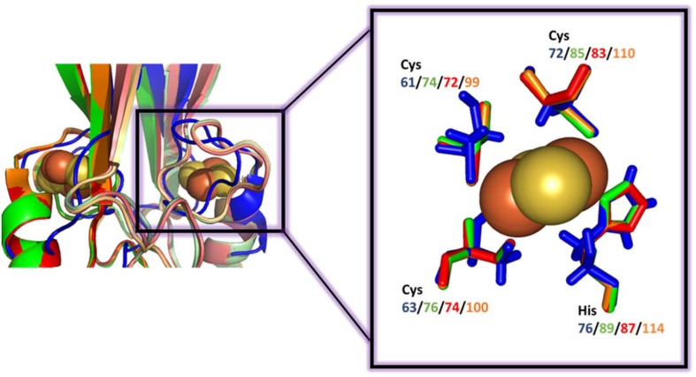
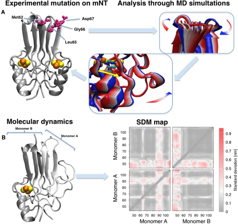
NEET proteins comprise a new class of [2Fe-2S] cluster proteins. In human, three genes encode for NEET proteins: cisd1 encodes mitoNEET (mNT), cisd2 encodes the Nutrient-deprivation autophagy factor-1 (NAF-1) and cisd3 encodes MiNT (Miner2). These recently discovered proteins play key roles in many processes related to normal metabolism and disease. Indeed, NEET proteins are involved in iron, Fe-S, and reactive oxygen homeostasis in cells and play an important role in regulating apoptosis and autophagy. mNT and NAF-1 are homodimeric and reside on the outer mitochondrial membrane. NAF-1 also resides in the membranes of the ER associated mitochondrial membranes (MAM) and the ER. MiNT is a monomer with distinct asymmetry in the molecular surfaces surrounding the clusters. Unlike its paralogs mNT and NAF-1, it resides within the mitochondria. NAF-1 and mNT share similar backbone folds to the plant homodimeric NEET protein (At-NEET), while MiNT's backbone fold resembles a bacterial MiNT protein. Despite the variation of amino acid composition among these proteins, all NEET proteins retained their unique CDGSH domain harboring their unique 3Cys:1His [2Fe-2S] cluster coordination through evolution. The coordinating exposed His was shown to convey the lability to the NEET proteins' [2Fe-2S] clusters. In this minireview, we discuss the NEET fold and its structural elements. Special attention is given to the unique lability of the NEETs' [2Fe-2S] cluster and the implication of the latter to the NEET proteins' cellular and systemic function in health and disease.

Keywords: Cisd(1–3) encoded NEET proteins; Iron-sulfur clusters; NEET-cluster lability; NEET-fold; [2Fe-2S].


Figures

Fig. 1
Fig. 1
NEET proteins CDGSH organization. The location of the CDGSH domain(s) is shown in (red box) bacterial MiNT (blue), At-NEET (green), mitoNEET (red) and NAF-1 (brown). Different textures of the boxes were used to distinguish between different domains: in-organelle domain (checker texture), inter-membrane domain (diagonal lines pattern) and cytosolic domain (full color). The sequence interval is reported for each domain. The different regions specified here are based on the sequence of each protein
Fig. 2
Fig. 2
NEET proteins’ structures solved by X-ray crystallography. Structure of monomeric bacterial MiNT (blue colored; PDB-ID: 3tbn [14]), and dimeric (monomers A are reported with lighter colors) At-NEET (green, 3s2q [30]), mitoNEET (red, 2qh7 [15]) and NAF-1 (brown, 4oo7 [38]) proteins, and their superposition. The [2Fe-2S] cluster atoms are shown in orange-yellow spheres. The superposition of the four proteins shows the high structural similarity shared between the NEET proteins. The crystallized part of the homodimeric NEET proteins is limited to the cytosolic domain (fully colored part in Fig. 1) without the linkers to the membrane and the intra-organelle parts
Fig. 3
Fig. 3
Central hydrophobic domains of the NEET proteins. The amino acids belonging to the hydrophobic central patch of mNT (red), NAF-1 (orange) and At-NEET (green) are shown in ball and stick representations over the structures of mNT [25], NAF-1 [38] and At-NEET [30] structures colored in grey shades from the brighter to the darker, respectively. The localization of the conserved Tyr is affected by the displacement of the surrounding amino acids, modulating, therefore, its position and orientation
Fig. 4
Fig. 4
Electrostatic potential on the NEET protein’s surface. The electrostatic potential values (estimated using NEET proteins’ force files [85] and APBS electrostatic [103]) of mNT [25], NAF-1 [38] and At-NEET [30] are here reported over each protein surface. The side facing the plane of the β-sheet (top) and the side view (bottom) are here reported. The color code refers to the electrostatic potential values reported on the right
Fig. 5
Fig. 5
[2Fe-2S] cluster-binding domain of NEET proteins. Comparison of the superposition of the cluster-binding domain using the same color-code as in Fig. 2. The details of the superposition of the [2Fe-2S] 3Cys:1His pocket of each protein is shown within the box. In the left hand side figure the overlap between the proteins is not ideal for MiNT. Nevertheless, the similarity of the inner coordination sphere of ligands of the different NEET proteins is high
Fig. 6
Fig. 6
mNT modifications and theoretical analysis. (a, left) Crystal structure of mNT [25] highlighting the allosteric mutated residues at the top of the β-cap (violet). All crystal structures are available of the mutated proteins [51]. The β-cap mutations alter the coordinated motions of the domain (a, center panel), correlated with the flexibility of the cluster binding domain (right) and, in particular, with the coordinating histidine (yellow colored arrow) [51]. The colors of the cartoon structure in the central and right panels span from blue to red representing the movement along the principal vibrational mode of the protein. (b, left) Representative structure of the mNT in absence of one [2Fe-2S] cluster from monomer A obtained using replica exchange molecular dynamics [52]. (b, right) The effects of the cluster absence on standard deviation maps [52]. Here each pixel represents the standard deviation of the distance between each residue couple. The regions which are mostly affected by the cluster absence are the α-helix of the monomer losing the cluster and the L1 domain of the other [85]

References

    1. Beinert H, Holm RH, Munck E. Science. 1997;277:653–659. - PubMed
    1. Balk J, Lobreaux S. Trends Plant Sci. 2005;10:324–331. - PubMed
    1. Rees DC, Howard JB. Curr Opin Chem Biol. 2000;4:559–566. - PubMed
    1. Beinert H. J Biol Inorg Chem. 2000;5:2–15. - PubMed
    1. Lill R. Nature. 2009;460:831–838. - PubMed

Publication types

Substances

LinkOut - more resources