

A step towards stereotactic navigation during pelvic surgery: 3D nerve topography
- PMID: 29435745
- PMCID: PMC6061054
- DOI: 10.1007/s00464-018-6086-3
A step towards stereotactic navigation during pelvic surgery: 3D nerve topography
Abstract
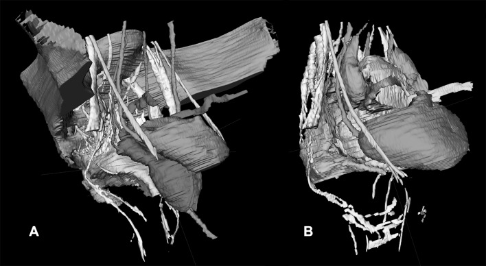
Background: Long-term morbidity after multimodal treatment for rectal cancer is suggested to be mainly made up by nerve-injury-related dysfunctions. Stereotactic navigation for rectal surgery was shown to be feasible and will be facilitated by highlighting structures at risk of iatrogenic damage. The aim of this study was to investigate the ability to make a 3D map of the pelvic nerves with magnetic resonance imaging (MRI).
Methods: A systematic review was performed to identify a main positional reference for each pelvic nerve and plexus. The nerves were manually delineated in 20 volunteers who were scanned with a 3-T MRI. The nerve identifiability rate and the likelihood of nerve identification correctness were determined.
Results: The analysis included 61 studies on pelvic nerve anatomy. A main positional reference was defined for each nerve. On MRI, the sacral nerves, the lumbosacral plexus, and the obturator nerve could be identified bilaterally in all volunteers. The sympathetic trunk could be identified in 19 of 20 volunteers bilaterally (95%). The superior hypogastric plexus, the hypogastric nerve, and the inferior hypogastric plexus could be identified bilaterally in 14 (70%), 16 (80%), and 14 (70%) of the 20 volunteers, respectively. The pudendal nerve could be identified in 17 (85%) volunteers on the right side and in 13 (65%) volunteers on the left side. The levator ani nerve could be identified in only a few volunteers. Except for the levator ani nerve, the radiologist and the anatomist agreed that the delineated nerve depicted the correct nerve in 100% of the cases.
Conclusion: Pelvic nerves at risk of injury are usually visible on high-resolution MRI with dedicated scanning protocols. A specific knowledge of their course and its application in stereotactic navigation is suggested to improve quality of life by decreasing the likelihood of nerve injury.
Keywords: Anatomy; Autonomic nervous system; Hypogastric plexus; Magnetic resonance imaging; Medical; Neuronavigation; Topography.
Conflict of interest statement
Dr. L. Soler receives remuneration (personal fees) from Visible Patient S. A. S. though he has no direct conflicts of interest with content discussed in this manuscript. Drs. A. G. F. Melani receives remuneration (payment for services not otherwise identified as salary such as consulting fees, honoraria) from Medtronic, Ethicon, Intuitive Surgical and Verb Surgical though he has no direct conflicts of interest with content discussed in this manuscript. Prof. J. Marescaux is president of both IRCAD and IHU Strasbourg, which are partly funded by Karl Storz, Medtronic, and Siemens Healthcare though he has no direct conflict of interest with content discussed in this manuscript. Dr. A. R. Wijsmuller, Dr. C. Giraudeau, Dr. J. Leroy, Dr. G. J. Kleinrensink, Dr. E Rociu, Drs. L. G. Romagnolo, Dr. V. Agnus, Dr. M. Diana, Drs. B. Dallemagne, and Dr. D. Mutter have no conflicts of interest or financial ties to disclose.
Figures


References
-
- Wallner C, et al. Causes of fecal and urinary incontinence after total mesorectal excision for rectal cancer based on cadaveric surgery: a study from the Cooperative Clinical Investigators of the Dutch total mesorectal excision trial. J Clin Oncol. 2008;26(27):4466–4472. doi: 10.1200/JCO.2008.17.3062. - DOI - PubMed
Publication types
MeSH terms
LinkOut - more resources
Full Text Sources
Other Literature Sources
Medical
