

Alterations in Gene and Protein Expression of Cannabinoid CB2 and GPR55 Receptors in the Dorsolateral Prefrontal Cortex of Suicide Victims
- PMID: 29435814
- PMCID: PMC6095782
- DOI: 10.1007/s13311-018-0610-y
Alterations in Gene and Protein Expression of Cannabinoid CB2 and GPR55 Receptors in the Dorsolateral Prefrontal Cortex of Suicide Victims
Abstract
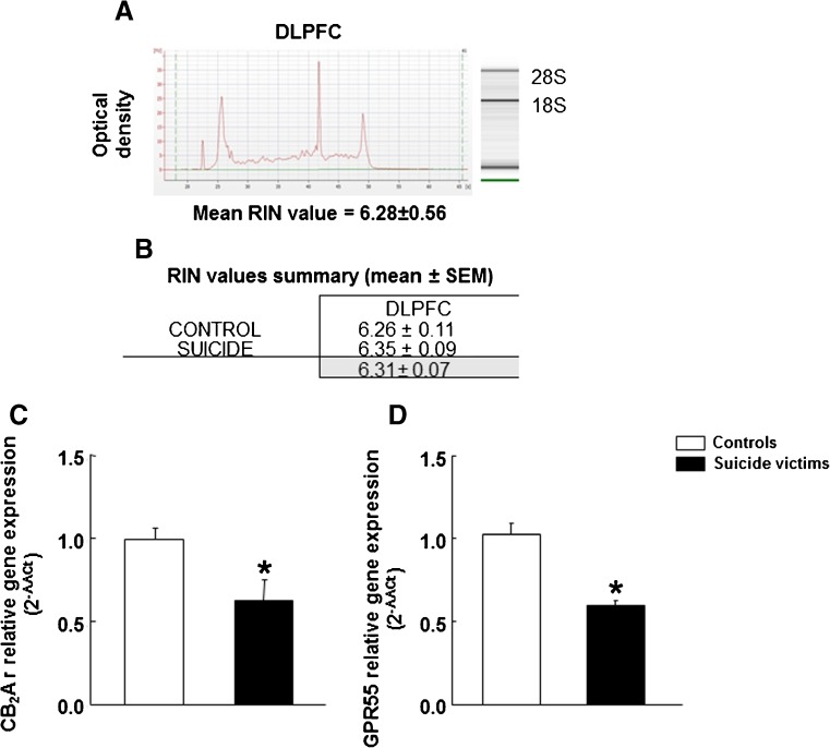
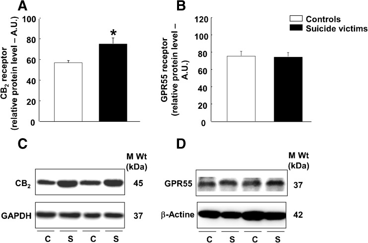
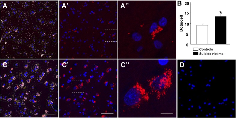
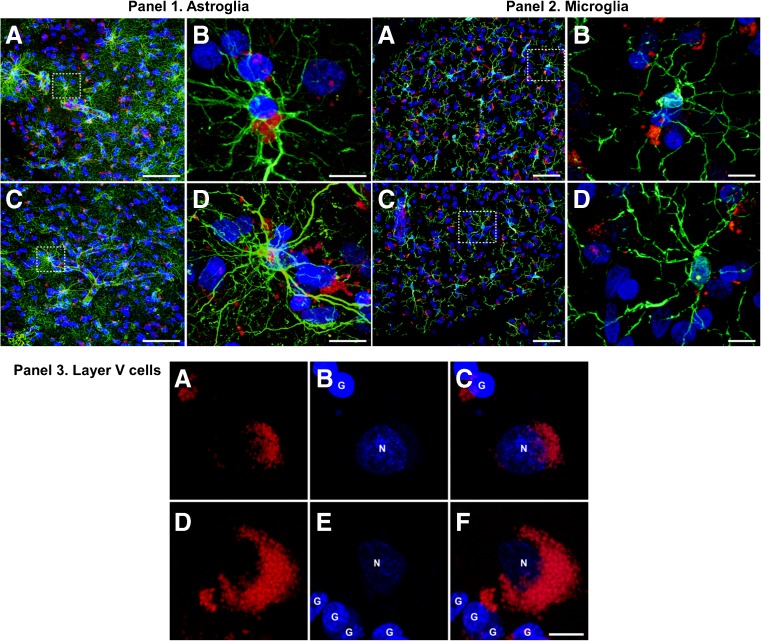
Recent studies point to the cannabinoid CB2 receptors (CB2r) and the non-cannabinoid receptor GPR55 as potential key targets involved in the response to stress, anxiety, and depression. Considering the close relationship between neuropsychiatric disorders and suicide, the purpose of this study was to evaluate the potential alterations of CB2r and GPR55 in suicide victims. We analyzed gene and protein expression of both receptors by real-time PCR and western blot, respectively, in the dorsolateral prefrontal cortex (DLPFC) of 18 suicide victims with no clinical psychiatric history or treatment with anxiolytics or antidepressants, and 15 corresponding controls. We used in situ proximity ligation assay to evaluate whether the receptors formed heteromeric complexes and to determine the expression level of these heteromers, also assessing the co-expression of heteromers in neurons, astroglia, or microglia cells. CB2r and GPR55 gene expressions were significantly lower (by 33 and 41%, respectively) in the DLPFC of suicide cases. CB2r protein expression was higher, as were CB2-GPR55 heteroreceptor complexes. The results also revealed the presence of CB2-GPR55 receptor heteromers in both neurons and astrocytes, whereas microglial cells showed no expression. We did not observe any significant alterations of GPR55 protein expression. Additional studies will be necessary to evaluate if these alterations are reproducible in suicide victims diagnosed with different psychiatric disorders. Taken together, the results suggest that CB2r and GPR55 may play a relevant role in the neurobiology of suicide.
Keywords: CB2r; Dorsolateral prefrontal cortex; GPR55; Gene expression; In situ proximity ligation assay; Suicide.
Conflict of interest statement
The authors declare that they have no conflicts of interest.
Figures
Similar articles
-
Heteromers Formed by GPR55 and Either Cannabinoid CB1 or CB2 Receptors Are Upregulated in the Prefrontal Cortex of Multiple Sclerosis Patients.Int J Mol Sci. 2024 Apr 10;25(8):4176. doi: 10.3390/ijms25084176. Int J Mol Sci. 2024. PMID: 38673761 Free PMC article.
-
Expression of GPR55 and either cannabinoid CB1 or CB2 heteroreceptor complexes in the caudate, putamen, and accumbens nuclei of control, parkinsonian, and dyskinetic non-human primates.Brain Struct Funct. 2020 Sep;225(7):2153-2164. doi: 10.1007/s00429-020-02116-4. Epub 2020 Jul 20. Brain Struct Funct. 2020. PMID: 32691218
-
Targeting CB2-GPR55 receptor heteromers modulates cancer cell signaling.J Biol Chem. 2014 Aug 8;289(32):21960-72. doi: 10.1074/jbc.M114.561761. Epub 2014 Jun 18. J Biol Chem. 2014. PMID: 24942731 Free PMC article.
-
New blood brothers: the GPR55 and CB2 partnership.Cell Res. 2011 Oct;21(10):1391-2. doi: 10.1038/cr.2011.77. Epub 2011 May 3. Cell Res. 2011. PMID: 21537344 Free PMC article. Review. No abstract available.
-
Progress in brain cannabinoid CB2 receptor research: From genes to behavior.Neurosci Biobehav Rev. 2019 Mar;98:208-220. doi: 10.1016/j.neubiorev.2018.12.026. Epub 2019 Jan 3. Neurosci Biobehav Rev. 2019. PMID: 30611802 Free PMC article. Review.
Cited by
-
Chronic Pain and Psychiatric Conditions.Complex Psychiatry. 2022 Sep 15;9(1-4):24-43. doi: 10.1159/000527041. eCollection 2023 Jan-Dec. Complex Psychiatry. 2022. PMID: 37034825 Free PMC article. Review.
-
Beta-Caryophyllene, a Cannabinoid Receptor Type 2 Selective Agonist, in Emotional and Cognitive Disorders.Int J Mol Sci. 2024 Mar 11;25(6):3203. doi: 10.3390/ijms25063203. Int J Mol Sci. 2024. PMID: 38542177 Free PMC article. Review.
-
Modulation of neuroinflammation and oxidative stress by targeting GPR55 - new approaches in the treatment of psychiatric disorders.Mol Psychiatry. 2024 Dec;29(12):3779-3788. doi: 10.1038/s41380-024-02614-5. Epub 2024 May 25. Mol Psychiatry. 2024. PMID: 38796643 Free PMC article. Review.
-
Endocannabinoid-mediated neuromodulation in the main olfactory bulb at the interface of environmental stimuli and central neural processing.Eur J Neurosci. 2022 Feb;55(4):1002-1014. doi: 10.1111/ejn.15186. Epub 2021 Apr 2. Eur J Neurosci. 2022. PMID: 33724578 Free PMC article. Review.
-
Cannabinoid CB1 Receptor Involvement in the Actions of CBD on Anxiety and Coping Behaviors in Mice.Pharmaceuticals (Basel). 2022 Apr 13;15(4):473. doi: 10.3390/ph15040473. Pharmaceuticals (Basel). 2022. PMID: 35455470 Free PMC article.
References
Publication types
MeSH terms
Substances
Grants and funding
- RETICS16/0017/0014/Ministry of Science and Innovation, Spain/International
- BFU2012-37907/Ministry of Science and Innovation, Spain/International
- PNSD 2015IO16, FIS PI14/00438/Ministry of Health, Spain/International
- 2014-SGR-1236/Agència de Gestió d'Ajuts Universitaris i de Recerca/International
- 201413-30/Fundació la Marató de TV3/International
LinkOut - more resources
Full Text Sources
Other Literature Sources
Medical
