Review
. 2018 May;7(3):e312.
doi: 10.1002/wdev.312. Epub 2018 Feb 13.

The zebrafish: A fintastic model for hematopoietic development and disease

Affiliations
Review

The zebrafish: A fintastic model for hematopoietic development and disease

Aniket V Gore et al. Wiley Interdiscip Rev Dev Biol. 2018 May.

Abstract

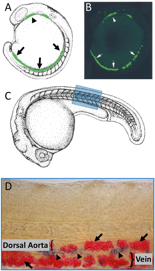
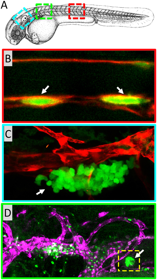
Hematopoiesis is a complex process with a variety of different signaling pathways influencing every step of blood cell formation from the earliest precursors to final differentiated blood cell types. Formation of blood cells is crucial for survival. Blood cells carry oxygen, promote organ development and protect organs in different pathological conditions. Hematopoietic stem and progenitor cells (HSPCs) are responsible for generating all adult differentiated blood cells. Defects in HSPCs or their downstream lineages can lead to anemia and other hematological disorders including leukemia. The zebrafish has recently emerged as a powerful vertebrate model system to study hematopoiesis. The developmental processes and molecular mechanisms involved in zebrafish hematopoiesis are conserved with higher vertebrates, and the genetic and experimental accessibility of the fish and the optical transparency of its embryos and larvae make it ideal for in vivo analysis of hematopoietic development. Defects in zebrafish hematopoiesis reliably phenocopy human blood disorders, making it a highly attractive model system to screen small molecules to design therapeutic strategies. In this review, we summarize the key developmental processes and molecular mechanisms of zebrafish hematopoiesis. We also discuss recent findings highlighting the strengths of zebrafish as a model system for drug discovery against hematopoietic disorders. This article is categorized under: Adult Stem Cells, Tissue Renewal, and Regeneration > Stem Cell Differentiation and Reversion Vertebrate Organogenesis > Musculoskeletal and Vascular Nervous System Development > Vertebrates: Regional Development Comparative Development and Evolution > Organ System Comparisons Between Species.

Keywords: blood; blood precursors; hemangioblasts; hematopoiesis; hematopoietic disorders; hematopoietic stem cells; leukemia; zebrafish.


Figures

Figure 1:
Figure 1:
Primitive hematopoiesis in zebrafish is responsible for generating the first populations of erythroid and myeloid cells. (A) Camera lucida drawing with superimposed green coloring depicting expression of an ETS transcription factor in the anterior (arrowhead) and posterior (arrows) lateral plate mesoderm. Primitive blood lineages are specified in anterior and posterior lateral plate mesoderm during early somitogenenesis. (B) Confocal image of a 16 hour-old Tg(fli1a:egfp)y1 embryo showing transgenic expression of EGFP in the anterior (arrowhead) and posterior (arrows) lateral plate mesoderm. (C) Camera lucida drawing of a 31 hpf zebrafish embryo with a blue box noting the approximate region of the trunk shown in panel D. (D) Double in situ hybridization staining for hbae1.1 primitive erythrocytes (red, arrows) and cmyb developing HSPCs (blue, arrowheads) in the ventral floor of the dorsal aorta of 32 hpf embryo.
Figure 2:
Figure 2:
Definitive hematopoiesis in zebrafish is responsible for generating all types of blood cells. (A) Camera lucida drawing of a 48 hpf zebrafish embryo with red, blue, and green colored boxes noting the approximate regions of the embryo shown in panels B, C, and D, respectively. (B) HSPCs develop in the ventral wall of the dorsal aorta. Confocal image of the mid trunk of a 32 hpf Tg(runx1+23:EGFP); Tg(kdrl:mCherry-caax) double-transgenic embryo showing EGFP-positive developing HSPCs (green; arrows) in the ventral floor of the mCherry-positive dorsal aorta (red). (C) The developing zebrafish thymus. Lateral view of a 5 dpf Tg(lck:GFP); Tg(kdrl:mCherry) double-transgenic embryo, just ventral to the otolith, showing GFP-positive thymocytes (green; arrow) developing adjacent to an mCherry-positive blood vessel (red). (D) The zebrafish head kidney is a major organ of definitive hematopoiesis equivalent to the bone marrow of mammals. Confocal image of the from the anterior trunk of a 5 dpf Tg(runx1+23:EGFP); Tg(lyve1:dsRed) double-transgenic embryo, showing GFP-positive hematopoietic cells (green; yellow box, white arrow) developing adjacent to an mCherry-positive vein (purple).
Figure 3:
Figure 3:
Schematic diagram illustrating the different sites of zebrafish hematopoiesis used throughout development. Stage of development is indicated in hours post fertilization (hpf) or days post fertilization (dpf). RBI, rostral blood islands; PLM, posterior lateral-plate mesoderm; ICM, intermediate cell mass; DA, dorsal aorta; CHT, caudal hematopoietic tissue.

References

    1. Paik EJ, Zon LI. Hematopoietic development in the zebrafish. Int J Dev Biol. 2010;54(6–7):1127–37. - PubMed
    1. Ciau-Uitz A, Monteiro R, Kirmizitas A, Patient R. Developmental hematopoiesis: ontogeny, genetic programming and conservation. Exp Hematol. 2014;42(8):669–83. - PubMed
    1. Tamplin OJ, Durand EM, Carr LA, Childs SJ, Hagedorn EJ, Li P, et al. Hematopoietic stem cell arrival triggers dynamic remodeling of the perivascular niche. Cell. 2015;160(1–2):241–52. - PMC - PubMed
    1. Hoggatt J, Singh P, Sampath J, Pelus LM. Prostaglandin E2 enhances hematopoietic stem cell homing, survival, and proliferation. Blood. 2009;113(22):5444–55. - PMC - PubMed
    1. North TE, Goessling W, Walkley CR, Lengerke C, Kopani KR, Lord AM, et al. Prostaglandin E2 regulates vertebrate haematopoietic stem cell homeostasis. Nature. 2007;447(7147):1007–11. - PMC - PubMed

Publication types