

Diagnosis and management of cerebral venous thrombosis
- PMID: 29436443
- PMCID: PMC6330914
- DOI: 10.7861/clinmedicine.18-1-75
Diagnosis and management of cerebral venous thrombosis
Erratum in
-
Editorial note.Clin Med (Lond). 2018 Mar;18(2):192. doi: 10.7861/clinmedicine.18-2-192a. Clin Med (Lond). 2018. PMID: 29626040 Free PMC article. No abstract available.
Abstract
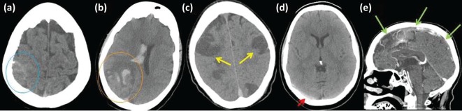
Cerebral venous thrombosis (CVT) is rare and accounts for 0.5% of all strokes. Its clinical presentation is variable and diagnosis requires a high index of clinical suspicion in conjunction with neuroradiological diagnostic support. Treatment options are limited and are mostly based on consensus. Therefore, familiarity with international guidelines is important. Outcome is often good and most patients make a full recovery, although a small proportion suffers death or disability. Here, we describe the clinical features, risk factors, acute imaging features, management and complications of CVT.
Keywords: anticoagulation; cerebral venography and heparin; cerebral venous sinus thrombosis; cerebral venous thrombosis; imaging; stroke.
© Royal College of Physicians 2018. All rights reserved.
Figures
References
-
- Bousser M-G. Ferro JM. Cerebral venous thrombosis: an update. Lancet Neurol. 2007;6:162–70. - PubMed
-
- Coutinho JM. Zuurbier SM. Aramideh M. Stam J. The incidence of cerebral venous thrombosis: a cross-sectional study. Stroke. 2012;43:3375–7. - PubMed
-
- Ferro JM. Canhao P. Cerebral venous sinus thrombosis: update on diagnosis and management. Curr Cardiol Rep. 2014;16:523. - PubMed
-
- Saposnik G. Barinagarrementeria F. Brown RD, et al. Diagnosis and management of cerebral venous thrombosis: a statement for healthcare professionals from the American Heart Association/American Stroke Association. Stroke. 2011;42:1158–92. - PubMed
-
- Bousser M-G. Russell RWR. Cerebral venous thrombosis. London:: Saunders; 1997.
Publication types
MeSH terms
LinkOut - more resources
Full Text Sources
Other Literature Sources
