Review
. 2018 Feb 7;82(1):e00057-17.
doi: 10.1128/MMBR.00057-17. Print 2018 Jun.

The Ontogeny of a Neutrophil: Mechanisms of Granulopoiesis and Homeostasis

Affiliations
Review

The Ontogeny of a Neutrophil: Mechanisms of Granulopoiesis and Homeostasis

Shelley M Lawrence et al. Microbiol Mol Biol Rev. .

Abstract

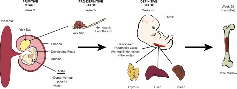
Comprising the majority of leukocytes in humans, neutrophils are the first immune cells to respond to inflammatory or infectious etiologies and are crucial participants in the proper functioning of both innate and adaptive immune responses. From their initial appearance in the liver, thymus, and spleen at around the eighth week of human gestation to their generation in large numbers in the bone marrow at the end of term gestation, the differentiation of the pluripotent hematopoietic stem cell into a mature, segmented neutrophil is a highly controlled process where the transcriptional regulators C/EBP-α and C/EBP-ε play a vital role. Recent advances in neutrophil biology have clarified the life cycle of these cells and revealed striking differences between neonatal and adult neutrophils based on fetal maturation and environmental factors. Here we detail neutrophil ontogeny, granulopoiesis, and neutrophil homeostasis and highlight important differences between neonatal and adult neutrophil populations.

Keywords: apoptosis; cell death; chemokine; degranulation; extracellular traps; granulopoiesis; hematopoiesis; innate immunity; neonates; neutrophils; phagocytosis.


Figures

FIG 1
FIG 1
Ontogeny of hematopoiesis. The origin of human blood cells begins in the mesoderm of the extraembryonic yolk sac around the third week of embryogenesis and is known as the primitive stage. The emergence of blood cells from the hemogenic endothelium begins within the yolk sac in week 5 during the prodefinitive (second) stage and is regulated by dorsal-ventral polarity and Notch signaling. Blood cell production then transitions to the ventral wall of the aorta at around the seventh to eighth week during the definitive stage and is regulated by the transcription factor Runx1 in a process known as the endothelial-to-hematopoietic transition (EHT), where HSCs are first identified. After the eighth week of gestation, early blood cells seed the liver, thymus, and spleen until the seventh month of gestation, when hematopoiesis transitions to the bone marrow. After this time, the bone marrow becomes the sole site for platelet and red and white blood cell formation.
FIG 2
FIG 2
Differentiation of common myeloid progenitor cells. Once destined to become a myeloid cell, the HSC enters a well-described, closely regulated process that results in the development of both megakaryocyte/erythroid and granulocyte/macrophage lineages from a pluripotent common myeloid progenitor cell. Whereas C/EBP-α and PU.1 induce CMPs to differentiate into monocytes and macrophages, C/EBP-ε and Gfi-1 generate neutrophils and eosinophils. It is the acetylation of C/EBP-ε at specific lysines (K121 and K198) and the lack of expression of GATA-1, however, that cause early CMPs to ultimately differentiate into neutrophils instead of eosinophils.
FIG 3
FIG 3
Granulopoiesis and associated transcription factors. Terminal neutrophil maturation is characterized by the sequential formation of the three different neutrophil granules and secretory vesicles as well as nuclear segmentation. Granulopoiesis begins with the development of azurophilic granules in myeloblasts and early promyelocytes and ends after the creation of secretory vesicles in mature, segmented cells. Neutrophil granule formation is hierarchical and dependent upon the timing of constituent protein biosynthesis, while exocytosis occurs in the reverse but ordered sequence. Gene expressions of GATA-1, C/EBP-ζ, AML-1, and c-Myc are imperative for azurophilic granule formation. The creation of specific granules occurs in conjunction with declining AML-1, c-Myc, and CDP concentrations. Reductions in the levels of CDP relieve its repression of C/EBP-ε genes, such as gp91phox, allowing the C/EBP-ε-induced transcription of both C/EBP-δ and specific granule proteins. Once the neutrophil matures into a metamyelocyte, it can no longer proliferate, marking the beginning of terminal neutrophil differentiation. This change results from the inhibition of proliferative genes, AML-1, C/EBP-γ, and CDP, and the emergence of antiproliferative factors such as C/EBP-δ and C/EBP-ζ. The transcription factor C/EBP-ε becomes downregulated as gene expressions for C/EBP-β, C/EBP-δ, and C/EBP-ζ are enhanced to form gelatinase granules.

References

    1. Kennedy AD, DeLeo FR. 2009. Neutrophil apoptosis and the resolution of infection. Immunol Res 43:25–61. doi: 10.1007/s12026-008-8049-6. - DOI - PubMed
    1. Medvinsky A, Rybtsov S, Taoudi S. 2011. Embryonic origin of the adult hematopoietic system: advances and questions. Development 138:1017–1031. doi: 10.1242/dev.040998. - DOI - PubMed
    1. Lacaud G, Kouskoff V. 2017. Hemangioblast, hemogenic endothelium, and primitive versus definitive hematopoiesis. Exp Hematol 49:19–24. doi: 10.1016/j.exphem.2016.12.009. - DOI - PubMed
    1. Tavian M, Peault B. 2005. Embryonic development of the human hematopoietic system. Int J Dev Biol 49:243–250. doi: 10.1387/ijdb.041957mt. - DOI - PubMed
    1. Tober J, Koniski A, McGrath KE, Vemishetti R, Emerson R, de Mesy-Bentley KK, Waugh R, Palis J. 2007. The megakaryocyte lineage originates from hemangioblast precursors and is an integral component both of primitive and of definitive hematopoiesis. Blood 109:1433–1441. doi: 10.1182/blood-2006-06-031898. - DOI - PMC - PubMed

MeSH terms

LinkOut - more resources