. 2018 Apr;17(4):5878-5886.
doi: 10.3892/mmr.2018.8556. Epub 2018 Feb 2.

Effects of bradykinin on TGF‑β1‑induced epithelial‑mesenchymal transition in ARPE‑19 cells

Affiliations

Effects of bradykinin on TGF‑β1‑induced epithelial‑mesenchymal transition in ARPE‑19 cells

Qingquan Wei et al. Mol Med Rep. 2018 Apr.

Abstract

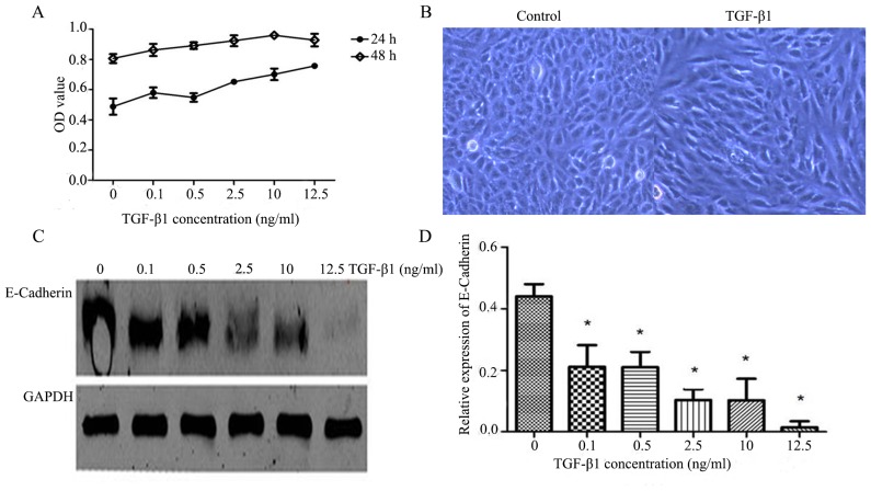
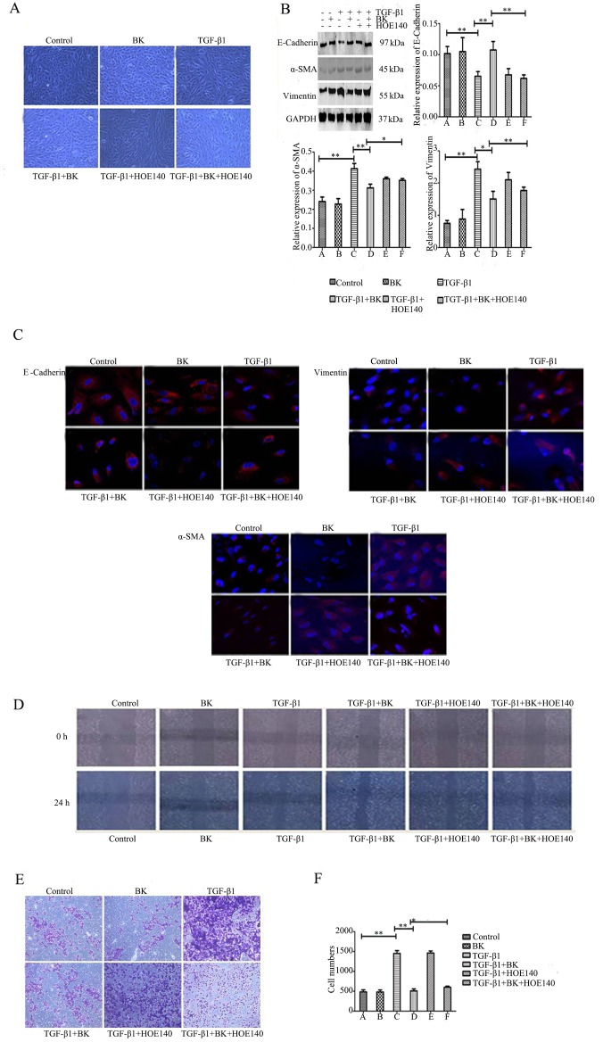
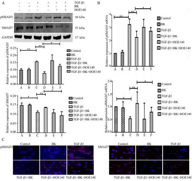
The aim of the present study was to investigate the effects of bradykinin (BK) on an epithelial-mesenchymal transition (EMT) model in retinal pigment epithelium (RPE) cells through exposure to transforming growth factor‑β1 (TGF‑β1). The aim was to improve the effect of BK on proliferative vitreoretinopathy (PVR) progression, and to find a novel method of clinical prevention and treatment for PVR. The morphology of ARPE‑19 cells was observed using an inverted phase‑contrast microscope. A Cell Counting Kit‑8 was used to assess the effects of TGF‑β1 on the proliferation of ARPE‑19 cells. Western blotting and immunofluorescence were used to detect the expression levels of the epithelial marker E‑cadherin, mesenchymal markers α‑smooth muscle actin (SMA) and vimentin, and phosphorylated (p) mothers against decapentaplegic homolog (Smad)3 and Smad7 of the TGF/Smad signaling pathway. Wound healing tests and Transwell assays were performed to detect cell migration ability. Reverse transcription‑quantitative polymerase chain reaction (RT‑qPCR) analysis was performed to detect the expression levels of pSmad3 and Smad7 in the TGF/Smad signaling pathway. The results revealed that the addition of 10 ng/ml TGF‑β1 resulted in the expression of factors associated with EMT in ARPE‑19 cells. BK decreased the expression levels of the mesenchymal markers α‑SMA and vimentin, and increased the expression of the epithelial marker E‑cadherin. BK decreased cell migration in TGF‑β1‑induced EMT. These effects were reversed by HOE‑140, a specific BK 2 receptor antagonist. BK significantly downregulated the expression of pSmad3 and upregulated the expression of Smad7 in TGF‑β1‑treated ARPE‑19 cells, and the protective alterations produced by BK were inhibited by HOE‑140. In conclusion, 10 ng/ml TGF‑β1 resulted in EMT in ARPE‑19 cells and BK served a negative role in TGF‑β1‑induced EMT. BK had effects in TGF‑β1‑induced EMT by upregulating the expression of Smad7 and downregulating the expression of pSmad3 in TGF‑β/Smad signaling pathway, indicating that BK may be a novel and effective therapy for PVR.

Keywords: bradykinin; roliferative vitreoretinopathy; transforming growth factor-β1; epithelial-mesenchymal transition; retinal pigment epithelium.


Figures

Figure 1.
Figure 1.
Treatment with 10 ng/ml TGF-β1 for 48 h results in epithelial-mesenchymal transition-associated features. (A) Different concentrations of TGF-β1 affected the proliferation of ARPE-19 cells. (B) Morphological alterations were detected between the control group and the TGF-β1 group using an inverted phase-contrast microscope (magnification, ×100). The optimal concentration of TGF-β1 was 10 ng/ml and the optimal incubation time was 48 h. (C) Western blot analysis was used to detect the expression levels of E-cadherin with different concentrations of TGF-β1, and (D) the results were quantified. All experiments were performed in triplicate. *P<0.05 vs. control. TGF-β1, transforming growth factor-β1; OD, optical density.
Figure 2.
Figure 2.
BK inhibits TGF-β1-induced epithelial-mesenchymal transitionin ARPE-19 cells, and a B2R inhibitor attenuates the role of BK. ARPE-19 cells were treated with BK, TGF-β1, BK plus TGF-β1, TGF-β1 plus HOE140, BK plus TGF-β1 plus HOE140, and Dulbecco's modified Eagle's medium for 48 h prior todetection. (A) Morphological alterations were detected using an inverted phase-contrast microscope (magnification, ×100). (B) Western blot analysis detected the expression levels of E-cadherin, α-SMA and vimentin in each group. (C) The protein expression of E-cadherin, α-SMA and vimentin was identified by immunofluorescence staining (magnification, ×200). (D) A wound healing assay tested the cell migration of a scratched edge. (E) Cells that had migrated through the Transwell chamber filter over 18 h were stained with crystal violet, and imaged at ×50 magnification. (F) The number of migrated ARPE-19 cells in each group. **P<0.001 and *P<0.05. TGF-β1, transforming growth factor-β1; BK, bradykinin; α-SMA, α-smooth muscle actin.
Figure 3.
Figure 3.
BK inhibits TGF-β1-induced epithelial-mesenchymal transition via the TGF/Smad signaling pathway. ARPE-19 cells were treated with BK, TGF-β1, BK plus TGF-β1, TGF-β1 plus HOE140, BK plus TGF-β1 plus HOE140 and Dulbecco's modified Eagle's medium for 48 h before detection. (A) Protein expression of pSmad3 and Smad7 was identified by western blot analysis. (B) Reverse transcription-quantitative polymerase chain reaction analysis and (C) immunofluorescence staining (magnification, ×200). **P<0.001 and *P<0.05. TGF-β1, transforming growth factor-β1; BK, bradykinin; pSmad, phosphorylated mothers against decapentaplegic homolog.

Similar articles

Cited by

References

    1. Pennock S, Haddock LJ, Eliott D, Mukai S, Kazlauskas A. Is neutralizing vitreal growth factors aviable strategy to prevent proliferative vitreoretinopathy? Prog Retin Eye Res. 2014;40:16–34. doi: 10.1016/j.preteyeres.2013.12.006. - DOI - PubMed
    1. Umazume K, Tsukahara R, Liu L, Fernandez de Castro JP, McDonald K, Kaplan HJ, Tamiya S. Role of retinal pigment epithelial cell β-catenin signaling in experimental proliferative vitreoretinopathy. Am J Pathol. 2014;184:1419–1428. doi: 10.1016/j.ajpath.2014.01.022. - DOI - PubMed
    1. Tosi GM, Marigliani D, Romeo N, Toti P. Disease pathways in proliferative vitreoretinopathy: An ongoing challenge. J Cell Physiol. 2014;229:1577–1583. doi: 10.1002/jcp.24606. - DOI - PubMed
    1. Li H, Wang H, Wang F, Gu Q, Xu X. Snail involves in the transforming growth factor β1-mediated epithelial-mesenchymal transition of retinal pigment epithelial cells. PLoS One. 2011;6:e23322. doi: 10.1371/journal.pone.0023322. - DOI - PMC - PubMed
    1. Bartel DP. MicroRNAs: Target recognition and regulatory functions. Cell. 2009;136:215–233. doi: 10.1016/j.cell.2009.01.002. - DOI - PMC - PubMed

MeSH terms