. 2018 Apr;17(4):5860-5868.
doi: 10.3892/mmr.2018.8606. Epub 2018 Feb 13.

The effect of foxp3-overexpressing Treg cells on non-small cell lung cancer cells

Affiliations

The effect of foxp3-overexpressing Treg cells on non-small cell lung cancer cells

Jiangzhou Peng et al. Mol Med Rep. 2018 Apr.

Retraction in

Abstract

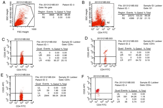
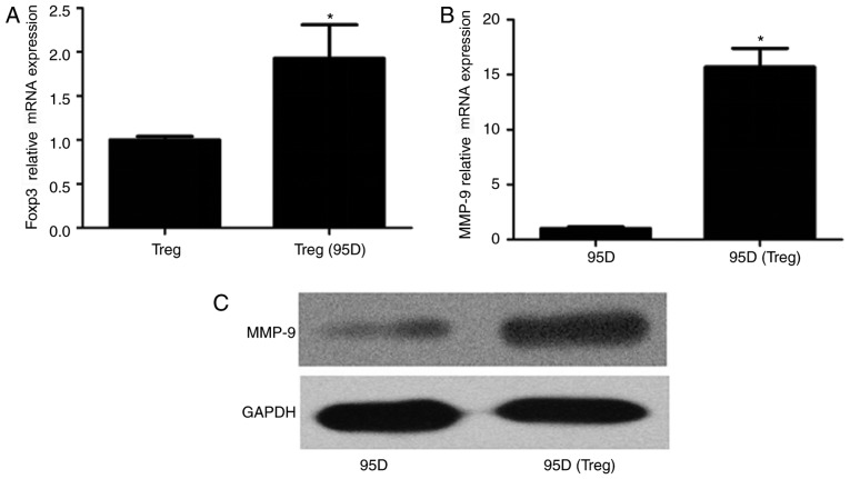
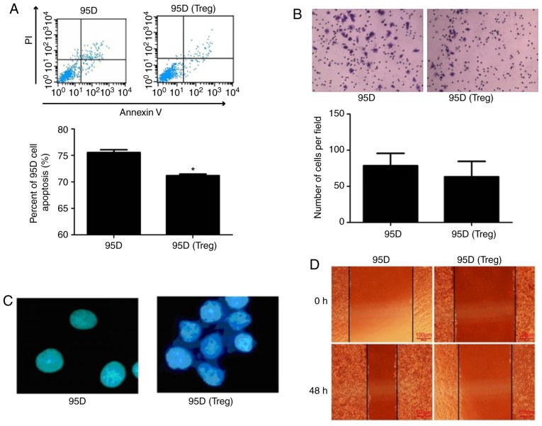
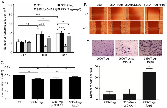
The aim of the present study was to investigate the novel mechanisms of forkhead box protein P3 (foxp3) in T regulatory (Treg) cells in lung cancer behavior. Treg cells were isolated from the peripheral blood of healthy volunteers and then co‑cultured with 95D cells. A plasmid overexpressing foxp3 was constructed and transfected into Treg cells and an MTS assay was performed to assess cell viability. Flow cytometry was performed to evaluate cell apoptosis and reverse transcription‑quantitative polymerase chain reaction was used to measure mRNA expression. A Transwell assay was used to assess cell invasion. Treg cells were successfully isolated from peripheral blood with purity of 94.26%. Foxp3 expression in Treg cells was significantly increased following co‑culture with 95D cells, while matrix metalloproteinase‑9 expression was upregulated in 95D cells co‑cultured with Treg cells. The apoptosis, invasion and migration abilities of 95D cells were suppressed by co‑culture with Treg cells, whereas the adhesive ability was enhanced. Foxp3 overexpression in Treg cells enhanced the viability and invasiveness of 95D cells, whereas cell adhesion and migration were decreased. The results of the present study demonstrate that the viability and invasiveness of 95D cells are enhanced by foxp3 overexpression in Treg cells, indicating that increased levels of foxp3 in the tumor microenvironment may promote tumor cell growth.

Keywords: T regulatory cell; non-small cell lung cancer; microenvironment; invasion; forkhead box protein P3.


Figures

Figure 1.
Figure 1.
Purity analysis of the isolated cells. Settings of the (A) T cell and (B) CD4 flow cytometer gates. (C) Ratio of CD4+CD25+ cells in CD4+ T cells. Sample: T cells. (D) Ratio of CD4+CD25+ cells in CD4+ T cells. Sample: CD4+CD127 cells. (E) Ratio of CD4+CD25+ cells in CD4+ T cells. Sample: CD25 cells. (F) Ratio of CD4+CD25+ cells in CD4+ T cells Sample: CD4+CD25+ cells. FSC, forward scatter; CD, cluster of differentiation; FITC, fluorescein isothiocyanate; UR, upper right; UL, upper left; LL, lower left; LR, lower right; APC, allophycocyanin; SSC, side scatter.
Figure 2.
Figure 2.
Gene expression in 95D cells following co-culture with Treg cells. (A) Foxp3 gene expression in Treg cells. *P<0.05 vs. Treg cells. (B) MMP-9 gene expression in 95D cells. *P<0.05 vs. 95D cells. (C) MMP-9 protein expression in 95D cells. Treg, regulatory T cells; foxp3, forkhead box protein P3; MMP, matrix metalloproteinase.
Figure 3.
Figure 3.
Impact of co-culture with Treg cells on the behavior of 95D cells. (A) Flow cytometry was used to assess cell apoptosis. (B) A Transwell assay was used to assess cell invasion (magnification, ×100). (C) Cell adhesion assay (magnification, ×400). (D) A wound healing assay was used to assess cell migration (magnification, ×100). *P<0.05 vs. 95D. PI, propidium iodide; Treg, regulatory T cells.
Figure 4.
Figure 4.
Effect of Foxp3 on cell co-culture. (A) Cell adhesion assay. (B) A wound healing assay was used to assess 95D cell migration. *P<0.05, **P<0.01, ***P<0.001. (C) MTS was used to assess 95D cell viability. *P<0.05. (D) A Transwell assay was used to assess 95D cell invasion. *P<0.05 vs. 95D+Treg and 95D+Treg-pcDNA3.1. OD, optical density; Treg, regulatory T cells; foxp3, forkhead box protein P3.

References

    1. Wang J, Jia Y, Zhao S, Zhang X, Wang X, Han X, Wang Y, Ma M, Shi J, Liu L. BIN1 reverses PD-L1-mediated immune escape by inactivating the c-MYC and EGFR/MAPK signaling pathways in non-small cell lung cancer. Oncogene. 2017;36:6235–6243. doi: 10.1038/onc.2017.217. - DOI - PubMed
    1. McGranahan N, Rosenthal R, Hiley CT, Rowan AJ, Watkins TBK, Wilson GA, Birkbak NJ, Veeriah S, Van Loo P, Herrero J, et al. Allele-Specific HLA loss and immune escape in lung cancer evolution. Cell. 2017;171:1259–1271.e11. doi: 10.1016/j.cell.2017.10.001. - DOI - PMC - PubMed
    1. Schafer CC, Wang Y, Hough KP, Sawant A, Grant SC, Thannickal VJ, Zmijewski J, Ponnazhagan S, Deshane JS. Indoleamine 2,3-dioxygenase regulates anti-tumor immunity in lung cancer by metabolic reprogramming of immune cells in the tumor microenvironment. Oncotarget. 2016;7:75407–75424. doi: 10.18632/oncotarget.12249. - DOI - PMC - PubMed
    1. Chan R, Sethi P, Jyoti A, McGarry R, Upreti M. Investigating the radioresistant properties of lung cancer stem cells in the context of the tumor microenvironment. Radiat Res. 2016;185:169–181. doi: 10.1667/RR14285.1. - DOI - PMC - PubMed
    1. Taylor JG, Gribben JG. Microenvironment abnormalities and lymphomagenesis: Immunological aspects. Semin Cancer Biol. 2015;34:36–45. doi: 10.1016/j.semcancer.2015.07.004. - DOI - PMC - PubMed

Publication types

MeSH terms

Substances