

Vertebro-vertebral fistula presenting as a pulsatile tinnitus
- PMID: 29437736
- PMCID: PMC5836622
- DOI: 10.1136/bcr-2017-222815
Vertebro-vertebral fistula presenting as a pulsatile tinnitus
Abstract
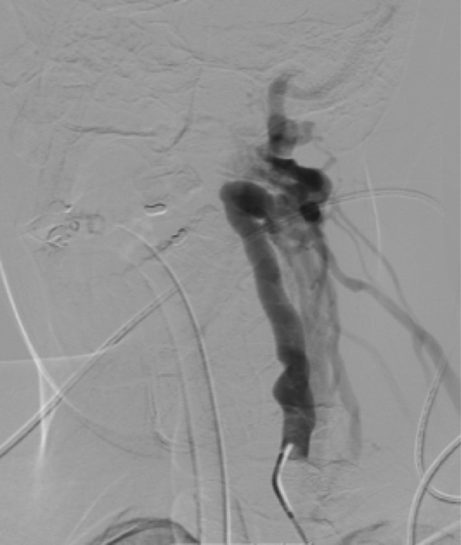
Tinnitus is the perception of sound in the absence of a corresponding external acoustic stimulus, resulting in an estimated prevalence of 10% to 15% in adults. Tinnitus may be classified as pulsatile (PT) or continuous (non-PT), and may be subjective (heard only by the patient) or objective (also audible to the examiner). PT is usually related to vascular causes and is pulse synchronous (coinciding with the patient's heartbeat). PT is much less common affecting approximately 4% of patients with tinnitus, but unlike non-PT, usually has a specific identifiable cause. We present a case of a man without previous otological disease or head trauma, with a left-ear subjective PT. MR angiography detected a left vertebro-vertebral arteriovenous fistula, which was treated by endovascular embolisation with important symptomatic relief.
Keywords: ear, nose and throat/otolaryngology; interventional radiology.
© BMJ Publishing Group Ltd (unless otherwise stated in the text of the article) 2018. All rights reserved. No commercial use is permitted unless otherwise expressly granted.
Conflict of interest statement
Competing interests: None declared.
Figures


References
Publication types
MeSH terms
LinkOut - more resources
Full Text Sources
Other Literature Sources
Medical