. 2018 Feb;11(2):e005852.
doi: 10.1161/CIRCEP.117.005852.

Altered Repolarization Reserve in Failing Rabbit Ventricular Myocytes: Calcium and β-Adrenergic Effects on Delayed- and Inward-Rectifier Potassium Currents

Affiliations

Altered Repolarization Reserve in Failing Rabbit Ventricular Myocytes: Calcium and β-Adrenergic Effects on Delayed- and Inward-Rectifier Potassium Currents

Bence Hegyi et al. Circ Arrhythm Electrophysiol. 2018 Feb.

Abstract

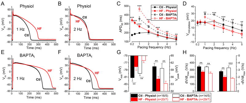
Background: Electrophysiological remodeling and increased susceptibility for cardiac arrhythmias are hallmarks of heart failure (HF). Ventricular action potential duration (APD) is typically prolonged in HF, with reduced repolarization reserve. However, underlying K+ current changes are often measured in nonphysiological conditions (voltage clamp, low pacing rates, cytosolic Ca2+ buffers).

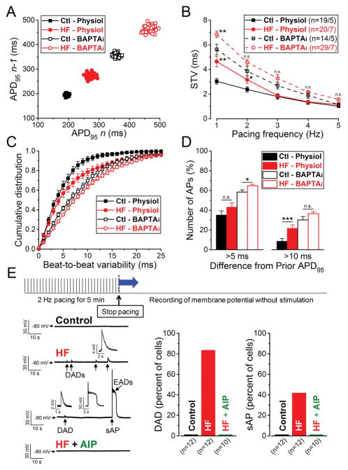
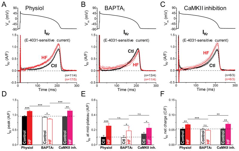
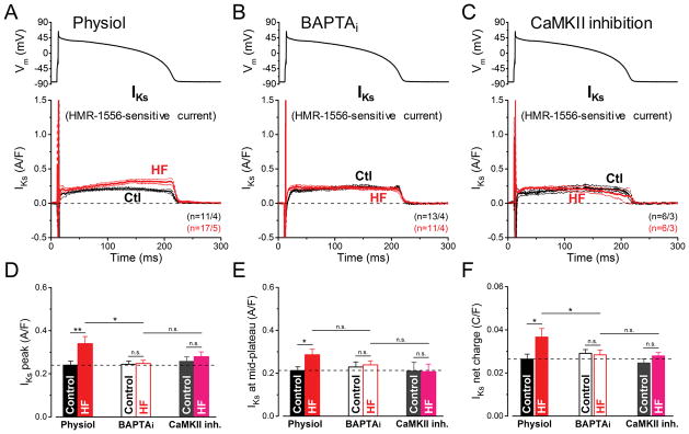
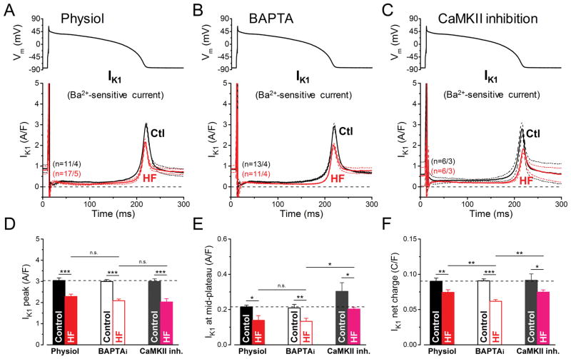
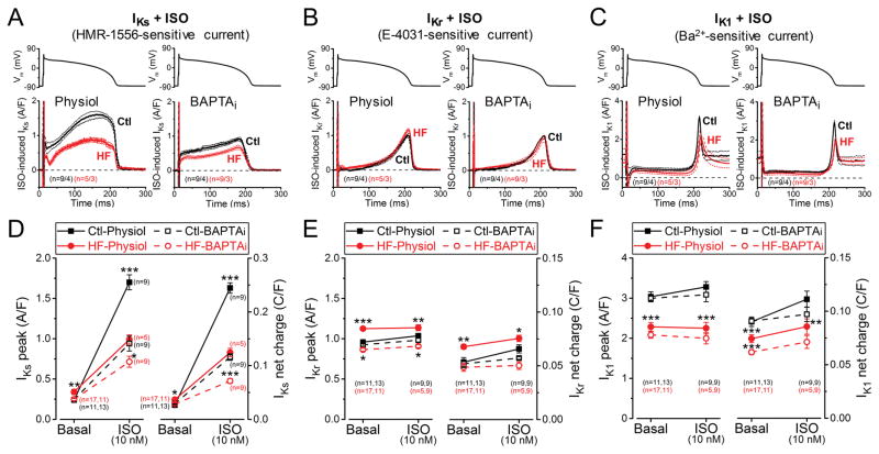
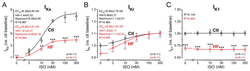
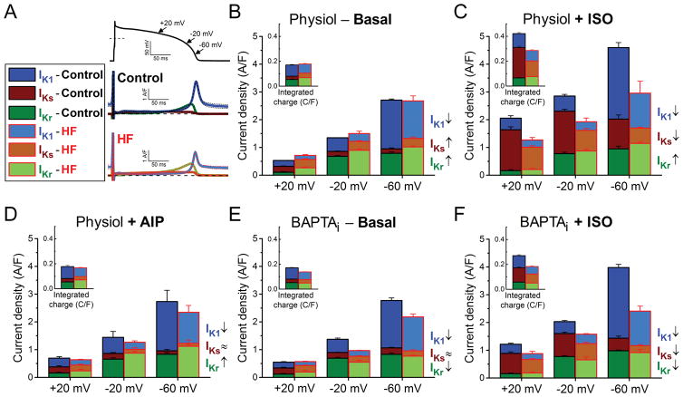
Methods and results: We measured the major K+ currents (IKr, IKs, and IK1) and their Ca2+- and β-adrenergic dependence in rabbit ventricular myocytes in chronic pressure/volume overload-induced HF (versus age-matched controls). APD was significantly prolonged only at lower pacing rates (0.2-1 Hz) in HF under physiological ionic conditions and temperature. However, when cytosolic Ca2+ was buffered, APD prolongation in HF was also significant at higher pacing rates. Beat-to-beat variability of APD was also significantly increased in HF. Both IKr and IKs were significantly upregulated in HF under action potential clamp, but only when cytosolic Ca2+ was not buffered. CaMKII (Ca2+/calmodulin-dependent protein kinase II) inhibition abolished IKs upregulation in HF, but it did not affect IKr. IKs response to β-adrenergic stimulation was also significantly diminished in HF. IK1 was also decreased in HF regardless of Ca2+ buffering, CaMKII inhibition, or β-adrenergic stimulation.

Conclusions: At baseline Ca2+-dependent upregulation of IKr and IKs in HF counterbalances the reduced IK1, maintaining repolarization reserve (especially at higher heart rates) in physiological conditions, unlike conditions of strong cytosolic Ca2+ buffering. However, under β-adrenergic stimulation, reduced IKs responsiveness severely limits integrated repolarizing K+ current and repolarization reserve in HF. This would increase arrhythmia propensity in HF, especially during adrenergic stress.

Keywords: action potential; calcium/calmodulin-dependent protein kinase II; electrophysiology; heart failure; potassium channels.


Figures

Figure 1
Figure 1. Frequency- and Ca2+-dependent changes of AP parameters in HF
(A–B) Representative action potentials (APs) recorded at 1 Hz and 2 Hz pacing in heart failure (HF) and healthy age-matched control cells with physiological solutions (Physiol). (C–D) Frequency dependence of AP duration measured at 95% of repolarization (APD95) and mid-plateau potential (Vmid-plateau). (E–F) Representative APs when cytosolic Ca2+ was buffered to nominally zero using 10 mM BAPTA in pipette solution (BAPTAi) at 1 Hz and 2 Hz pacing. (G) Resting membrane potential (Vrest) is less negative in HF in line with decreased AP peak (Vpeak) at 1 Hz. (H) Both maximal rate of rise (dV/dtmax) and maximal rate of phase 3 repolarization (-dV/dtmax) are significantly decreased in HF at 1 Hz. Columns and bars represent mean±SEM. n refers to cells/animals measured in each group. ANOVA with Bonferroni posttest, Ctl vs. HF, *p<0.05, **p<0.01, ***p<0.001.
Figure 2
Figure 2. Increased temporal variability of APD and arrhythmogenesis in HF
(A) Representative Poincare plots of 50 consecutive APD95 values at 1 Hz steady-state pacing in 4 individual myocytes from HF and age-matched control animals recorded with either preserved or buffered [Ca2+]i. (B) Frequency-dependent short-term variability of APD95 (STV). (C) Cumulative distribution curves of individual beat-to-beat variability values at 1 Hz pacing. (D) Percentage of total APs measured at 1 Hz pacing having beat-to-beat APD95 variability of >5 ms and >10 ms in consecutive beats. Columns and bars represent mean±SEM. n refers to cells/animals measured in each group. ANOVA with Bonferroni posttest, *p<0.05, **p<0.01, ***p<0.001. (E) Arrhythmogenic diastolic activities were tested using the pacing protocol shown above. (Left) Representative records in control, HF and AIP (CaMKII inhibitor, 1 μM) treated HF cells are shown below. HF cells frequently showed delayed afterdepolarizations (DADs, enlarged in insets) and spontaneous AP (sAP, enlarged in inset). Early afterdepolarizations (EADs, enlarged in inset) were also superimposed on the sAP repolarization. (Right) Percentage of cells having DAD and sAP. 10/12 cells and 5/12 cells showed DADs and sAP in HF, respectively, whereas no DAD, sAP or EAD was observed in control and following AIP treatment in HF.
Figure 3
Figure 3. Ca2+-dependent upregulation of IKr in HF under AP-clamp
The rapid component of delayed rectifier K+ current (IKr) was measured as E-4031 (1 μM)-sensitive current in HF and age-matched control cells. AP-clamp using a prerecorded typical AP (shown above) was applied at 2 Hz. (A) IKr traces (mean±SEM) recorded under preserved [Ca2+]i cycling (Physiol). IKr is not only increased in HF cells having [Ca2+]i transients, but it activates earlier during the AP. (B) IKr traces (mean±SEM) recorded under buffered [Ca2+]i using 10 mM BAPTA in the pipette (BAPTAi). Buffering [Ca2+]i significantly reduced IKr density in HF below the control level, but it had no effect in control. (C) IKr traces (mean±SEM) recorded in cells pretreated with the specific CaMKII inhibitor peptide AIP (1 μM). AIP had no effect on IKr both in control and in HF. (D) Peak IKr density is significantly upregulated in HF under AP by a Ca2+-dependent, but CaMKII-independent pathway. (E) IKr density measured at the mid-plateau increased in HF indicating earlier IKr accumulation during AP, which occurred independent of [Ca2+]i level and CaMKII activity. (F) Net charges carried by IKr in HF and age-matched control. Columns and bars represent mean±SEM. n refers to cells/animals measured in each group. ANOVA with Bonferroni posttest, *p<0.05, **p<0.01, ***p<0.001.
Figure 4
Figure 4. Ca2+/CaMKII-dependent upregulation of IKs in HF under AP-clamp
The slow component of delayed rectifier K+ current (IKs) was measured as HMR-1556 (1 μM)-sensitive current in HF and age-matched control cells. AP-clamp using a prerecorded typical AP (shown above) was applied at 2 Hz. (A) IKs traces (mean±SEM) recorded under preserved [Ca2+]i cycling (Physiol). Basal IKs is a tiny current under AP, yet it is significantly upregulated in HF cells having [Ca2+]i transients. (B) IKs traces (mean±SEM) recorded under buffered [Ca2+]i using 10 mM BAPTA in the pipette (BAPTAi). Buffering [Ca2+]i reduced IKs in HF back to its control level. (C) IKs traces (mean±SEM) recorded in cells pretreated with the specific CaMKII inhibitor peptide AIP (1 μM). AIP abolished the IKs upregulation in HF. (D) Peak IKs density is significantly upregulated in HF under AP by a Ca2+/CaMKII-dependent pathway. (E) IKs density measured at the mid-plateau of the AP. (F) Net charges carried by IKs in HF and age-matched control. Columns and bars represent mean±SEM. n refers to cells/animals measured in each group. ANOVA with Bonferroni posttest, *p<0.05, **p<0.01.
Figure 5
Figure 5. Reduction of IK1 in HF under AP-clamp
The inward rectifier K+ current (IK1) was measured as Ba2+ (100 μM)-sensitive current in HF and age-matched control cells. AP-clamp using a prerecorded typical AP (shown above) was applied at 2 Hz. (A–C) IK1 traces (mean±SEM) recorded under preserved [Ca2+]i cycling (Physiol), [Ca2+]i buffering (BAPTAi) and CaMKII inhibition using AIP. IK1 was significantly reduced in HF in all these conditions. (D–F) Statistics for peak IK1 density shows that the reduction is not influenced by [Ca2+]i buffering or acute CaMKII inhibition. (E) IK1 density measured at the mid-plateau of the AP was also reduced. (F) Net charges carried by IK1 was further reduced in HF when measured with [Ca2+]i buffering. Columns and bars represent mean±SEM. n refers to cells/animals measured in each group. ANOVA with Bonferroni posttest, *p<0.05, **p<0.01, ***p<0.001.
Figure 6
Figure 6. Altered response of K+ currents to β-AR stimulation in HF
IKs, IKr and IK1 currents were recorded following 2 min pretreatment with beta-adrenergic receptor agonist isoproterenol (ISO, 10 nM). (A) IKs traces (mean±SEM) under canonical AP-clamp at 2 Hz measured with preserved [Ca2+]i cycling (Physiol) and [Ca2+]i buffering using 10 mM BAPTA in the pipette (BAPTAi). (B) IKr traces (mean±SEM) under AP-clamp following ISO pretreatment in HF and age-matched control cells. (C) IK1 traces (mean±SEM) under AP-clamp following ISO pretreatment in physiological solutions and in BAPTAi. (D) Robust upregulation of IKs peak and net charge induced by ISO, which is reduced in BAPTAi indicating a Ca2+-dependent pathway in mediating the response of β-AR stimulation on IKs besides the classical PKA-dependent phosphorylation. HF cells showed significantly reduced IKs accumulation upon ISO application both with and without [Ca2+]i buffering. (E) IKr is slightly modulated by ISO both in control and HF. (F) IK1 peak density is not influenced by ISO, but IK1 net charge is slightly increased in HF due to altered rectification. Symbols and bars represent mean±SEM. n refers to cells measured in each group, and the cells in each group came from three to five individual animals. ANOVA with Bonferroni posttest, *p<0.05, **p<0.01, ***p<0.001.
Figure 7
Figure 7. β-AR responsiveness of K+ currents in HF
Dose-response effect of isoproterenol (ISO) on IKs, IKr and IK1 peak densities under AP-clamp at 2 Hz. Pipette solution contained 10 mM BAPTA. (A) IKs measured as HMR-sensitive current showed a robust accumulation following ISO application. IKS sensitivity (EC50) to ISO was unchanged in HF, whereas the response was significantly reduced in HF. (B) IKr was minimally affected by ISO treatment; however, neither the ISO-sensitivity nor the magnitude of the response were altered in HF. (C) IK1 peak density did not change following ISO application under buffered [Ca2+]i conditions and IK1 was uniformly decreased in HF. EC50 values, Hill coefficients and maximum responses were determined by fitting data to the Hill equation, indicated by solid lines. Symbols and bars represent mean±SEM. n refers to the number of cells measured in each group, and the cells in each group came from three to five individual animals. ANOVA with Bonferroni posttest, *p<0.05, **p<0.01, ***p<0.001.
Figure 8
Figure 8. Relative contribution of each K+ current to net repolarizing K+ current in HF
Relative contributions and magnitudes of the major repolarizing K+ currents (IKr, IKs, IK1) during AP are compared in different phases of the repolarization process in HF to those in age-matched control. (A) IKr, IKs and IK1 traces in control and HF measured under AP-clamp at 2 Hz without using any Ca2+ buffer or β-AR agonist. Mean traces and SEM are shown. (B) When [Ca2+]i cycling is preserved, upregulation of IKr and IKs compensates the decrease of IK1 in HF during phase 3 of AP. (C) Stimulation of β-ARs using isoproterenol (ISO, 10 nM) significantly upregulates IKs, thus ISO reverses IKr/IKs dominant pattern of repolarization during phase 3 of AP. However, HF cells are hyporesponsive to ISO-induced stimulation (i.e. IKs increases in a smaller extent than in control) thus the net repolarizing current is largely reduced in HF compared to control. Reversal in IKr/IKs dominance in repolarization following ISO treatment also fail to happen in HF. (D) CaMKII inhibition using the specific inhibitory peptide AIP abolishes the increase in IKs, whereas it does not affect IKr and IK1. (E) When [Ca2+]i is buffered (BAPTAi), the reduction in IK1 without the upregulation of IKr and IKs results in a significant decrease in the net repolarizing current in HF. (F) Under β-adrenergic stimulation in BAPTAi, HF cells show decrease in all three K+ currents compared to control. The contributions of these K+ currents to total net charge are shown in the insets. Columns and bars represent mean±SEM. Statistics and n numbers are shown in Figs. 3–6.

Comment in

References

    1. Tomaselli GF, Beuckelmann DJ, Calkins HG, Berger RD, Kessler PD, Lawrence JH, Kass D, Feldman AM, Marban E. Sudden cardiac death in heart failure. The role of abnormal repolarization. Circulation. 1994;90:2534–2539. - PubMed
    1. Watanabe E, Arakawa T, Uchiyama T, Tong M, Yasui K, Takeuchi H, Terasawa T, Kodama I, Hishida H. Prognostic significance of circadian variability of RR and QT intervals and QT dynamicity in patients with chronic heart failure. Heart Rhythm. 2007;4:999–1005. - PubMed
    1. Arsenos P, Gatzoulis KA, Dilaveris P, Gialernios T, Sideris S, Lazaros G, Archontakis S, Tsiachris D, Kartsagoulis E, Stefanadis C. The rate-corrected QT interval calculated from 24-hour holter recordings may serve as a significant arrhythmia risk stratifier in heart failure patients. Int J Cardiol. 2011;147:321–323. - PubMed
    1. Algra A, Tijssen JGP, Roelandt JRTC, Pool J, Lubsen J. QTc prolongation measured by standard 12-lead electrocardiography is an independent risk factor for sudden-death due to cardiac-arrest. Circulation. 1991;83:1888–1894. - PubMed
    1. Piccirillo G, Magri D, Matera S, Magnanti M, Torrini A, Pasquazzi E, Schifano E, Velitti S, Marigliano V, Quaglione R, Barilla F. QT variability strongly predicts sudden cardiac death in asymptomatic subjects with mild or moderate left ventricular systolic dysfunction: A prospective study. Eur Heart J. 2007;28:1344–1350. - PubMed

Publication types