. 2018 Mar 7;38(10):2569-2578.
doi: 10.1523/JNEUROSCI.1728-17.2018. Epub 2018 Feb 7.

Roles of Prefrontal Cortex and Mediodorsal Thalamus in Task Engagement and Behavioral Flexibility

Affiliations

Roles of Prefrontal Cortex and Mediodorsal Thalamus in Task Engagement and Behavioral Flexibility

Tobias F Marton et al. J Neurosci. .

Abstract

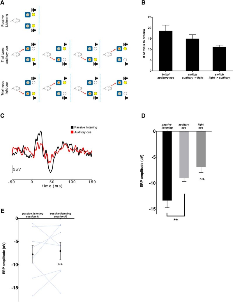
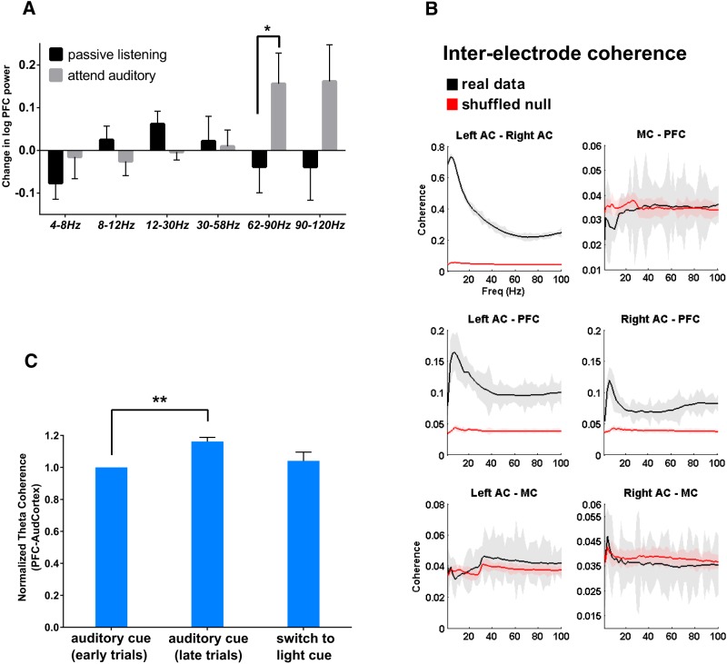
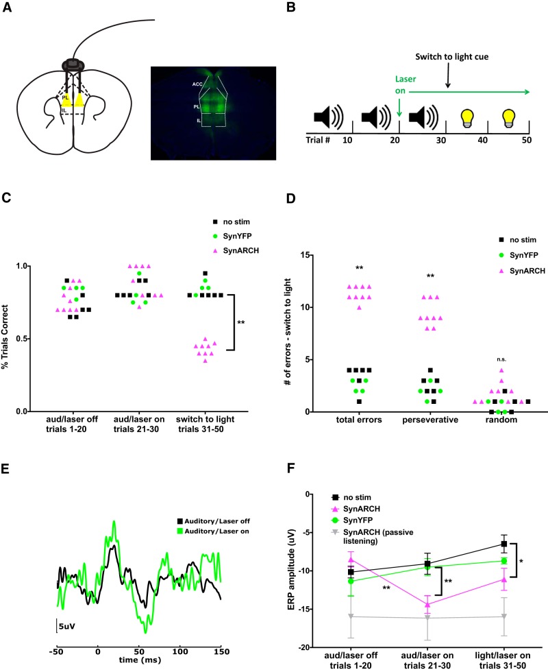
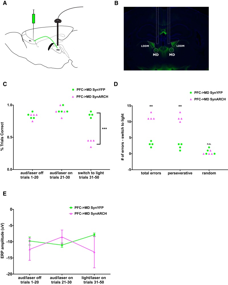
Behavioral tasks involving auditory cues activate inhibitory neurons within auditory cortex, leading to a reduction in the amplitude of auditory evoked response potentials (ERPs). One hypothesis is that this process, termed "task engagement," may enable context-dependent behaviors. Here we set out to determine (1) whether the medial prefrontal cortex (mPFC) plays a role in task engagement and (2) how task engagement relates to the context-dependent processing of auditory cues in male and female mice performing a decision-making task that can be guided by either auditory or visual cues. We found that, in addition to auditory ERP suppression, task engagement is associated with increased mPFC activity and an increase in theta band (4-7 Hz) synchronization between the mPFC and auditory cortex. Optogenetically inhibiting the mPFC eliminates the task engagement-induced auditory ERP suppression, while also preventing mice from switching between auditory and visual cue-based rules. However, mPFC inhibition, which eliminates task engagement-induced auditory ERP suppression, did not prevent mice from making decisions based on auditory cues. Furthermore, a more specific manipulation, selective disruption of mPFC outputs to the mediodorsal (MD) thalamus, is sufficient to prevent switching between auditory and visual rules but does not affect auditory ERPs. Based on these findings, we conclude that (1) the mPFC contributes to both task engagement and behavioral flexibility; (2) mPFC-MD projections are important for behavioral flexibility but not task engagement; and (3) task engagement, evidenced by the suppression of cortical responses to sensory input, is not required for sensory cue-guided decision making.SIGNIFICANCE STATEMENT When rodents perform choice-selection tasks based on sensory cues, neural responses to these cues are modulated compared with task-free conditions. Here we demonstrate that this phenomenon depends on the prefrontal cortex and thus represents a form of "top-down" regulation. However, we also show that this phenomenon is not critical for task performance, as rodents can make decisions based on specific sensory cues even when the task-dependent modulation of responses to those cues is abolished. Furthermore, disrupting one specific set of prefrontal outputs impairs rule switching but not the task-dependent modulation of sensory responses. These results show that the prefrontal cortex comprises multiple circuits that mediate dissociable functions related to behavioral flexibility and sensory processing.

Keywords: cognitive flexibility; decision making; evoked response potential; mediodorsal thalamus; prefrontal cortex.


Figures

Figure 1.
Figure 1.
Selective attention to auditory cues elicits suppression of the auditory ERP N1. A, Auditory–visual rule-switching task conditions. In passive listening, tone and light stimuli are presented, but neither tray contains a food reward. In the auditory rule condition, tones are played from the speaker on the same side as the rewarded food tray. The position of the light stimulus varies randomly between the rewarded (congruent with auditory stimulus) and unrewarded (incongruent with auditory stimulus) food trays, as shown in the four possible trial types. In the visual rule condition, once mice reach a criterion of 8 of 10 trials correct during the auditory rule portion of the task, the rewarded tray becomes paired with the light stimulus, and the location of the tones varies randomly between the rewarded (congruent) and unrewarded (incongruent) tray, as shown in the four possible trial types. B, Mice rapidly reached the performance criterion (8 of 10 trials correct) while shifting between auditory cue and light cue-based rules (n = 10). C, Auditory ERPs in one mouse generated from tones presented during passive listening (black) or the auditory rule condition (red) demonstrate suppression of the ERP N1 (negativity at 40–50 ms) during auditory cue-guided behavior. D, Quantification of the ERP N1 amplitude during passive listening versus the auditory rule versus visual rule task conditions (n = 12). **p < 0.01. E, ERP N1 amplitudes do not change during 2 consecutive days of passive listening to the auditory cue (no task engagement; n = 8).
Figure 2.
Figure 2.
mPFC gamma power and mPFC–AC theta coherence increase during auditory cue-guided behavior. A, mPFC power in the gamma band (60–120 Hz) increases during the auditory rule portion of the task compared with passive listening (n = 7). *p < 0.05. B, Coherence versus frequency for all electrode pairs (black) versus boot-strapped data sets (red). Gray shading indicates +/−1 SEM. Electrode pairs between PFC and AC show peak coherence in the theta (4–7 Hz) band. C, Theta coherence during early auditory rule trials (performance below 80% correct), late auditory rule trials (at least 8 of 10 trials correct), and visual rule trials (normalized to the coherence during the early attend auditory trails). There is a significant increase in mPFC–AC theta coherence as mice learn the auditory rule (n = 6). **p < 0.01.
Figure 3.
Figure 3.
Inhibiting mPFC potentiates the N1 of the auditory ERP and disrupts auditory–visual rule switching. A, AAV-Syn-ARCH3.0-eYFP was injected bilaterally into the mPFC. B, Schematic of the rule-switching task: mice first learned an auditory cue-based rule and then, after 30 trials, switched to a light cue-based rule. C, Optical inhibition of mPFC does not affect performance based on the established auditory rule but impairs switching to the new visual rule. **p < 0.01. D, Optical inhibition of mPFC results in perseverative errors during the rule switch (WT, n = 9; SynARCH, n = 6; YFP, n = 4). **p < 0.01. E, Example auditory ERPs from one mouse showing that optical inhibition of mPFC modulates sensory processing of the relevant cue by increasing the N1 component of the auditory ERP. F, Quantification of N1 amplitude across the three task conditions in the presence or absence of optogenetic inhibition. Optogenetic inhibition of mPFC increases the N1 amplitude of the auditory ERP, yielding an amplitude similar to that observed during passive (nonattended) listening (WT, n = 6; SynARCH, n = 6; YFP, n = 4). *p < 0.05, **p < 0.01.
Figure 4.
Figure 4.
Optogenetic inhibition of mPFC projections to MD thalamus disrupts rule switching but has no effect on auditory ERPs. A, AAV-Syn-ARCH3.0-eYFP was injected bilaterally into mPFC, and optical fibers were placed bilaterally over MD thalamus. PL = prelimbic cortex. B, Histology showing mPFC projection terminals expressing Syn-ARCH3.0-eYFP in the MD thalamus as well as in the nearby laterodorsal dorsomedial thalamus (LDDM). The location of optical fibers has been outlined. C, Optical inhibition of mPFC projections to MD does not affect performance based on an established auditory rule but disrupts performance during the switch to a visual rule (SynARCH, n = 4; YFP, n = 4). D, Optical inhibition of mPFC projections to MD results in perseverative errors during the attempt to switch to a visual rule. E, Quantification of the N1 amplitude of the auditory ERP across the three conditions. Optical inhibition of mPFC projections to MD had no significant effect on the N1 amplitude. ***p < 0.001; **p < 0.01.

References

    1. Amann LC, Gandal MJ, Halene TB, Ehrlichman RS, White SL, McCarren HS, Siegel SJ (2010) Mouse behavioral endophenotypes for schizophrenia. Brain Res Bull 83:147–161. 10.1016/j.brainresbull.2010.04.008 - DOI - PubMed
    1. Anticevic A, Haut K, Murray JD, Repovs G, Yang GJ, Diehl C, McEwen SC, Bearden CE, Addington J, Goodyear B, Cadenhead KS, Mirzakhanian H, Cornblatt BA, Olvet D, Mathalon DH, McGlashan TH, Perkins DO, Belger A, Seidman LJ, Tsuang MT, et al. (2015) Association of thalamic dysconnectivity and conversion to psychosis in youth and young adults at elevated clinical risk. JAMA Psychiatry 72:882–891. 10.1001/jamapsychiatry.2015.0566 - DOI - PMC - PubMed
    1. Birrell JM, Brown VJ (2000) Medial frontal cortex mediates perceptual attentional set shifting in the rat. J Neurosci 20:4320–4324. - PMC - PubMed
    1. Bissonette GB, Martins GJ, Franz TM, Harper ES, Schoenbaum G, Powell EM (2008) Double dissociation of the effects of medial and orbital prefrontal cortical lesions on attentional and affective shifts in mice. J Neurosci 28:11124–11130. 10.1523/JNEUROSCI.2820-08.2008 - DOI - PMC - PubMed
    1. Cardin JA, Carlén M, Meletis K, Knoblich U, Zhang F, Deisseroth K, Tsai LH, Moore CI (2009) Driving fast-spiking cells induces gamma rhythm and controls sensory responses. Nature 459:663–667. 10.1038/nature08002 - DOI - PMC - PubMed

Publication types