. 2018 Feb 6;9(1):e02287-17.
doi: 10.1128/mBio.02287-17.

Staphylococcus aureus HemX Modulates Glutamyl-tRNA Reductase Abundance To Regulate Heme Biosynthesis

Affiliations

Staphylococcus aureus HemX Modulates Glutamyl-tRNA Reductase Abundance To Regulate Heme Biosynthesis

Jacob E Choby et al. mBio. .

Abstract

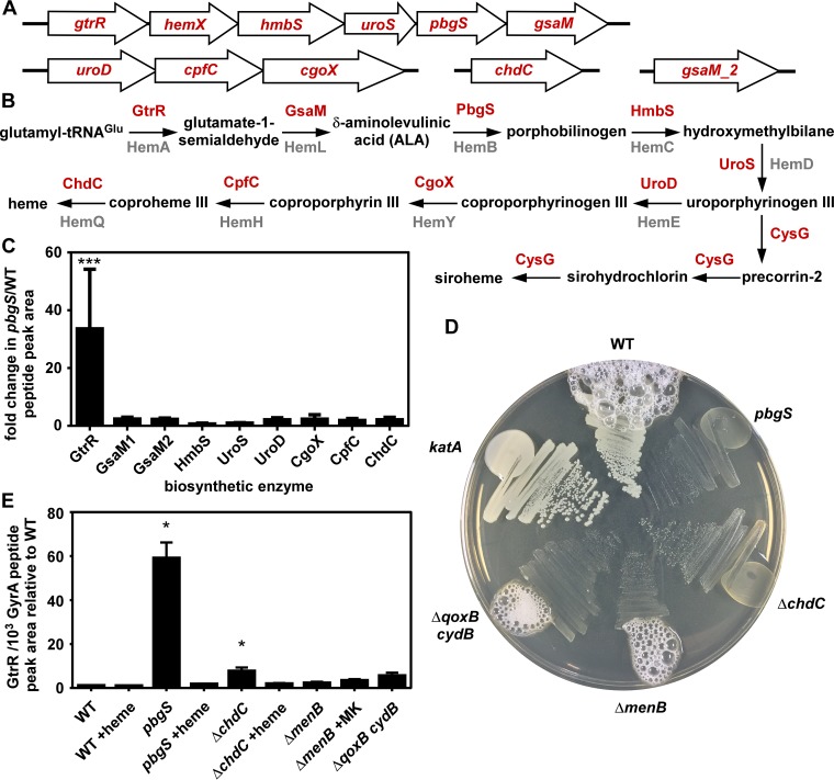
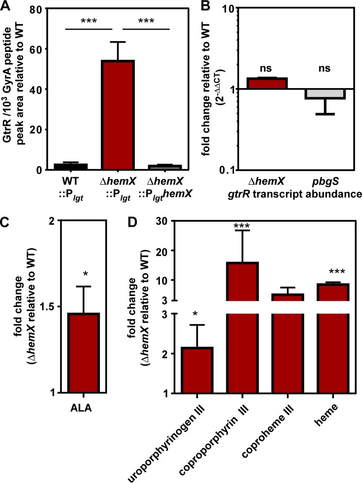
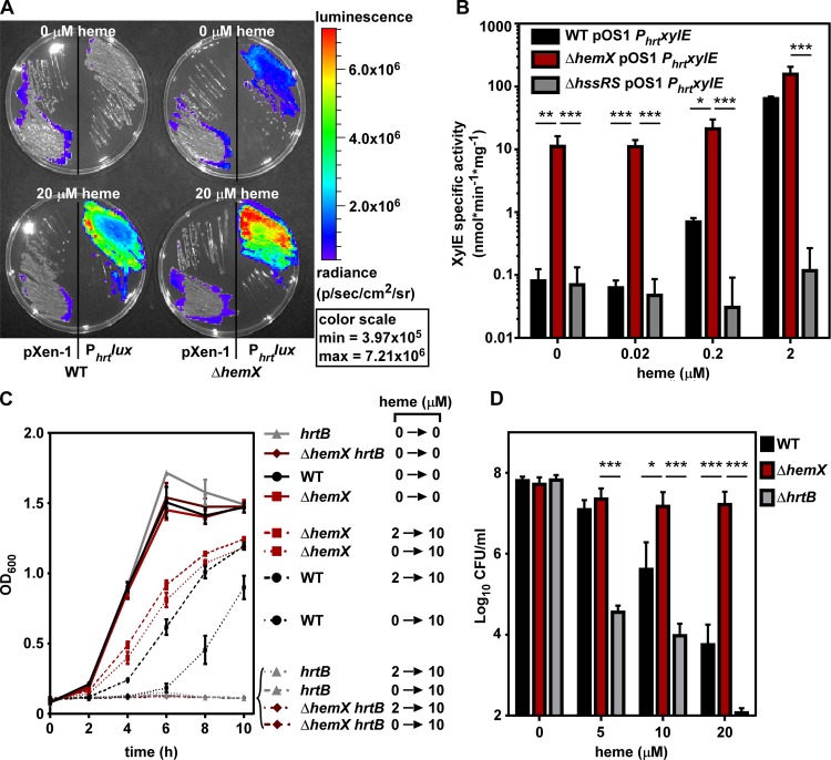
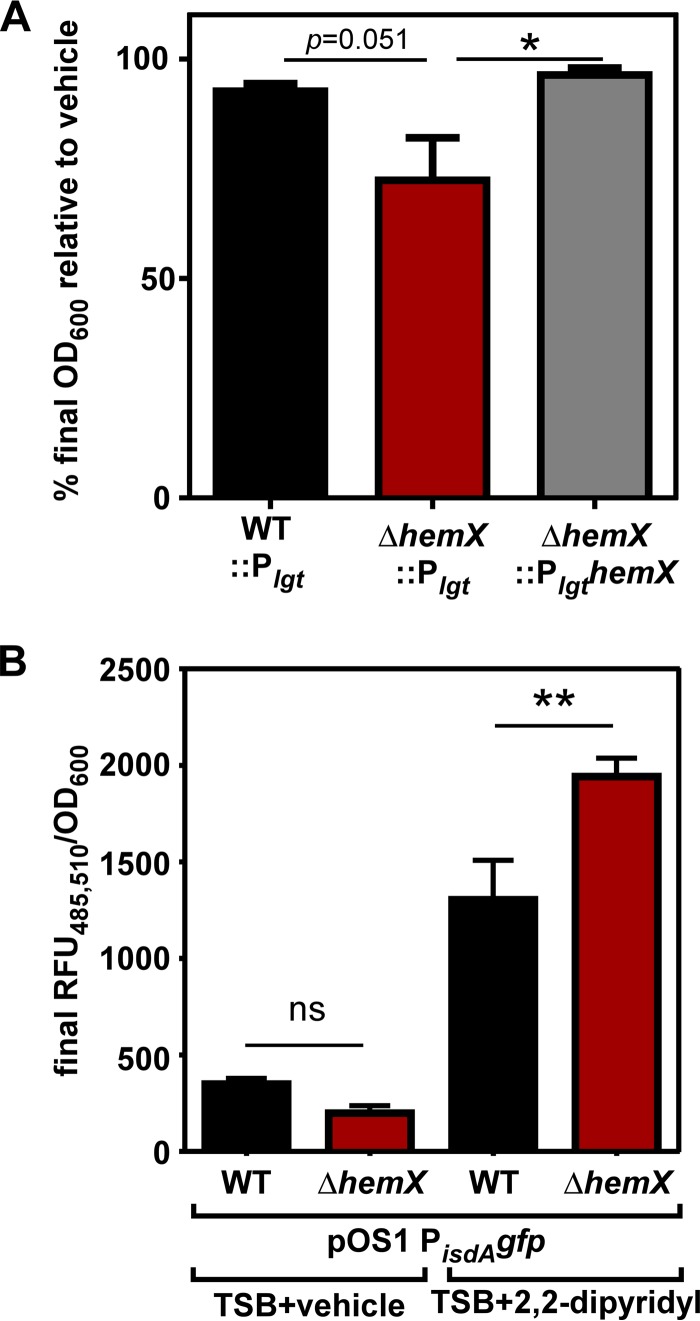
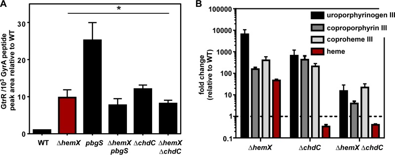
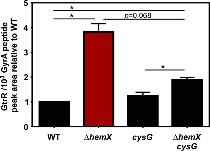
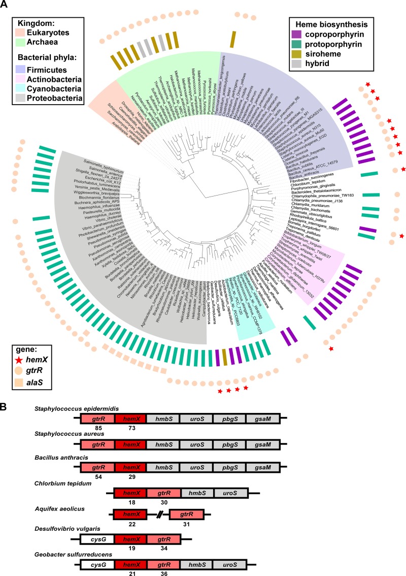
Staphylococcus aureus is responsible for a significant amount of devastating disease. Its ability to colonize the host and cause infection is supported by a variety of proteins that are dependent on the cofactor heme. Heme is a porphyrin used broadly across kingdoms and is synthesized de novo from common cellular precursors and iron. While heme is critical to bacterial physiology, it is also toxic in high concentrations, requiring that organisms encode regulatory processes to control heme homeostasis. In this work, we describe a posttranscriptional regulatory strategy in S. aureus heme biosynthesis. The first committed enzyme in the S. aureus heme biosynthetic pathway, glutamyl-tRNA reductase (GtrR), is regulated by heme abundance and the integral membrane protein HemX. GtrR abundance increases dramatically in response to heme deficiency, suggesting a mechanism by which S. aureus responds to the need to increase heme synthesis. Additionally, HemX is required to maintain low levels of GtrR in heme-proficient cells, and inactivation of hemX leads to increased heme synthesis. Excess heme synthesis in a ΔhemX mutant activates the staphylococcal heme stress response, suggesting that regulation of heme synthesis is critical to reduce self-imposed heme toxicity. Analysis of diverse organisms indicates that HemX is widely conserved among heme-synthesizing bacteria, suggesting that HemX is a common factor involved in the regulation of GtrR abundance. Together, this work demonstrates that S. aureus regulates heme synthesis by modulating GtrR abundance in response to heme deficiency and through the activity of the broadly conserved HemX.IMPORTANCEStaphylococcus aureus is a leading cause of skin and soft tissue infections, endocarditis, bacteremia, and osteomyelitis, making it a critical health care concern. Development of new antimicrobials against S. aureus requires knowledge of the physiology that supports this organism's pathogenesis. One component of staphylococcal physiology that contributes to growth and virulence is heme. Heme is a widely utilized cofactor that enables diverse chemical reactions across many enzyme families. S. aureus relies on many critical heme-dependent proteins and is sensitive to excess heme toxicity, suggesting S. aureus must maintain proper intracellular heme homeostasis. Because S. aureus provides heme for heme-dependent enzymes via synthesis from common precursors, we hypothesized that regulation of heme synthesis is one mechanism to maintain heme homeostasis. In this study, we identify that S. aureus posttranscriptionally regulates heme synthesis by restraining abundance of the first heme biosynthetic enzyme, GtrR, via heme and the broadly conserved membrane protein HemX.

Keywords: Staphylococcus aureus; heme; tetrapyrroles.


Figures

FIG 1
FIG 1
Heme deficiency increases GtrR abundance. (A) The genes encoding heme biosynthesis enzymes are located at four chromosomal loci. (B) An overview of the S. aureus heme and siroheme biosynthetic pathway. In red are the updated enzyme names set forth by Dailey and colleagues (14), which correspond to the previously used gene locus names in gray. (C) The abundance of each biosynthetic enzyme was measured by LC-MRM-MS/MS and quantified by integrated chromatogram peak areas. Graphed is the ratio of each enzyme’s abundance in a strain lacking pbgS relative to WT S. aureus; the data are the average from a single experiment performed in biological triplicate with standard deviation shown. Statistical significance was determined using a one-way analysis of variance (ANOVA) with Dunnett’s correction for multiple comparisons, using a reference value of 1.0. ***, P < 0.001. (D) The S. aureus strains listed were streaked onto rich agar medium plates, and after growth, hydrogen peroxide was added at the perimeter of each streak. (E) The abundance of GtrR was measured by LC-MRM-MS/MS in S. aureus strains treated with vehicle, heme, or menaquinone (MK). The data are the average from a single experiment performed in biological triplicate with standard deviation shown. Statistical significance was determined using a one-way ANOVA with Dunnett’s correction for multiple comparisons, comparing GtrR abundance for each condition relative to the WT. *, P < 0.05.
FIG 2
FIG 2
HemX regulates heme synthesis by maintaining low levels of GtrR in heme-proficient cells. (A) The abundance of GtrR was measured by LC-MRM-MS/MS in multiple S. aureus strains. The data are the average from a single experiment performed in biological triplicate with standard deviation shown. Statistical significance was determined using a one-way ANOVA with Dunnett’s correction for multiple comparisons, comparing GtrR abundance for each strain relative to the ΔhemX::Plgt strain. ***, P < 0.001. (B) Steady-state transcript abundance of gtrR mRNA isolated from mid-exponential growth of S. aureus strains was measured by qRT-PCR and is graphed as fold change relative to the WT. Data are combined from two independent experiments in biological triplicate with standard deviation shown. “ns” indicates no significance by one-way ANOVA with Dunnett’s correction for multiple comparisons, comparing fold change of the pbgS and ΔhemX strains to the WT. (C) δ-Aminolevulinic acid (ALA) abundance was measured in S. aureus strains by a spectrophotometric quantification. Graphed is the fold change of ALA in the ΔhemX mutant relative to the WT, with data combined from two independent experiments with three biological replicates with standard error of the mean shown. (D) Uroporphyrinogen III (detected as uroporphyin III), coproporphyrin III, coproheme III, and heme were quantified by LC-qTOF-MS. Graphed is the fold change of metabolite abundance in the ΔhemX mutant relative to the WT, from a single experiment performed in biological triplicate with standard error of the mean shown. For panels C and D, statistical significance was determined with Student’s t test comparing the ΔhemX mutant to the WT before data transformation to fold change. *, P < 0.05; ***, P < 0.0001.
FIG 3
FIG 3
Excess heme synthesis in the ΔhemX mutant activates the heme stress response. (A) Bioluminescence was imaged on agar medium plates containing vehicle or heme onto which strains were streaked. All four plates were imaged simultaneously, and luminescence was converted to a heat map with the scale shown on the right. (B) XylE catechol oxidase activity was measured in S. aureus strains after growth in vehicle or increasing concentrations of heme. The data are the average from three independent experiments each in biological triplicate with standard deviation shown. Statistical significance was determined using a two-way ANOVA with Tukey’s correction for multiple comparisons, comparing log-transformed data for the ΔhemX pOS1 PhrtxylE strain at each heme concentration to that of each other strain. *, P < 0.01; **, P < 0.001; ***, P < 0.0001. (C) Growth as measured by OD600 was monitored over time for S. aureus strains in medium containing either vehicle or 10 µM heme. Prior to the measured growth, the strains were pregrown to the stationary phase in medium containing vehicle or 2 µM heme. The data are the average of the means from at least three independent experiments each in biological triplicate with standard error of the mean shown. (D) Viable bacteria from S. aureus strains were enumerated after incubation for 2 h in medium containing vehicle or increasing amounts of heme. The data are the average of the means from three independent experiments each in biological triplicate with standard error of the mean shown. The y axis is set to the limit of detection. Statistical significance was determined using a two-way ANOVA with Tukey’s correction for multiple comparisons, comparing log-transformed data for the WT and ΔhrtB strains to the ΔhemX mutant at each heme concentration. *, P < 0.01; ***, P < 0.0001.
FIG 4
FIG 4
Unregulated heme synthesis alters iron homeostasis. (A) Growth was measured in minimal medium containing vehicle or 1 µM iron chelator EDDHA. Graphed is the final growth as measured by the OD600 for each S. aureus strain in medium containing EDDHA relative to vehicle. The data are the average of the means from five independent experiments each in at least biological triplicate with standard error of the mean shown. Statistical significance was determined using a one-way ANOVA with Dunnett’s correction for multiple comparisons, comparing the ΔhemX::Plgt strain to each other strain. *, P < 0.05. (B) The activity of the iron limitation-responsive promoter PisdA was measured by recording fluorescence intensity over time in rich medium containing vehicle or the iron chelator 2,2-dipyridyl. The data are the average of the means from three independent experiments each in biological triplicate with standard error of the mean shown. Statistical significance was determined using a one-way ANOVA with Sidak’s correction for multiple comparisons, comparing data for the WT and ΔhemX mutant under each condition. **, P < 0.01; ns, not significant.
FIG 5
FIG 5
Inactivation of HemX reduces GtrR abundance in heme-deficient strains. (A) The abundance of GtrR was measured by LC-MRM-MS/MS in multiple S. aureus strains. The data are the average from a single experiment performed in biological triplicate with standard deviation shown. Statistical significance was determined using a one-way ANOVA with Dunnett’s correction for multiple comparisons, comparing GtrR abundance for each strain relative to WT. *, P < 0.05. (B) Uroporphyrinogen III (detected as uroporphyin III), coproporphyrin III, coproheme III, and heme were quantified by LC-qTOF-MS. Graphed is the fold change of metabolite abundance in each mutant relative to the WT from a single experiment performed in biological triplicate with standard error of the mean shown.
FIG 6
FIG 6
Siroheme synthesis impacts GtrR levels under conditions of nitrite utilization. GtrR was measured by LC-MRM-MS/MS in multiple S. aureus strains grown anaerobically with NO3 provided as the terminal electron acceptor. The data are the average from a single experiment performed in biological triplicate with standard deviation shown. Statistical significance was determined using a one-way ANOVA with Dunnett’s correction for multiple comparisons, comparing GtrR abundance for each strain relative to the WT. *, P < 0.05.
FIG 7
FIG 7
hemX is conserved across bacterial phyla and invariably co-occurs (A) and colocalizes (B) with gtrR. (A) The occurrence of hemX (stars), gtrR (circles), and alaS (squares) homologues (outermost rings) was mapped onto the tree of life (53). The pathway by which protoheme is synthesized in each of the analyzed organisms is presented in the middle ring as follows (adapted from reference 14): the classic protoporphyrin-dependent pathway (teal), coproporphyrin-dependent path (purple), or siroheme-dependent path (gold). Gray rectangles mark the organisms that contain unusual combinations of genes normally involved in different pathways for protoheme synthesis (hybrid paths [14]). The absence of a rectangle in the middle ring indicates the absence of any known route for protoheme synthesis in an organism. Likewise, the absence of a circle (gtrR) or square (alaS) in the outermost ring shows the inability of an organism to produce tetrapyrroles of any kind. Note that hemX does not occur in such organisms. (B) The immediate genomic neighborhood of the hemX gene in seven representative genomes, with ClustalW alignment scores for HemX and GtrR for each organism relative to S. aureus.

References

    1. Choby JE, Skaar EP. 2016. Heme synthesis and acquisition in bacterial pathogens. J Mol Biol 428:3408–3428. doi: 10.1016/j.jmb.2016.03.018. - DOI - PMC - PubMed
    1. Klevens RM, Morrison MA, Nadle J, Petit S, Gershman K, Ray S, Harrison LH, Lynfield R, Dumyati G, Townes JM, Craig AS, Zell ER, Fosheim GE, McDougal LK, Carey RB, Fridkin SK, Active Bacterial Core surveillance (ABCs) MRSA Investigators . 2007. Invasive methicillin-resistant Staphylococcus aureus infections in the United States. JAMA 298:1763–1771. doi: 10.1001/jama.298.15.1763. - DOI - PubMed
    1. Hammer ND, Reniere ML, Cassat JE, Zhang Y, Hirsch AO, Hood MI, Skaar EP. 2013. Two heme-dependent terminal oxidases power Staphylococcus aureus organ-specific colonization of the vertebrate host. mBio 4:e00241-13. doi: 10.1128/mBio.00241-13. - DOI - PMC - PubMed
    1. Hammer ND, Schurig-Briccio LA, Gerdes SY, Gennis RB, Skaar EP. 2016. CtaM is required for menaquinol oxidase aa3 function in Staphylococcus aureus. mBio 7:e00823-16. doi: 10.1128/mBio.00823-16. - DOI - PMC - PubMed
    1. Dailey HA, Gerdes S, Dailey TA, Burch JS, Phillips JD. 2015. Noncanonical coproporphyrin-dependent bacterial heme biosynthesis pathway that does not use protoporphyrin. Proc Natl Acad Sci U S A 112:2210–2215. doi: 10.1073/pnas.1416285112. - DOI - PMC - PubMed

Publication types

MeSH terms

LinkOut - more resources