. 2018 Mar 28;92(8):e00146-18.
doi: 10.1128/JVI.00146-18. Print 2018 Apr 15.

A Polyamide Inhibits Replication of Vesicular Stomatitis Virus by Targeting RNA in the Nucleocapsid

Affiliations

A Polyamide Inhibits Replication of Vesicular Stomatitis Virus by Targeting RNA in the Nucleocapsid

Ryan H Gumpper et al. J Virol. .

Abstract

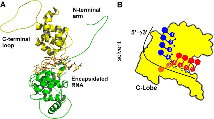
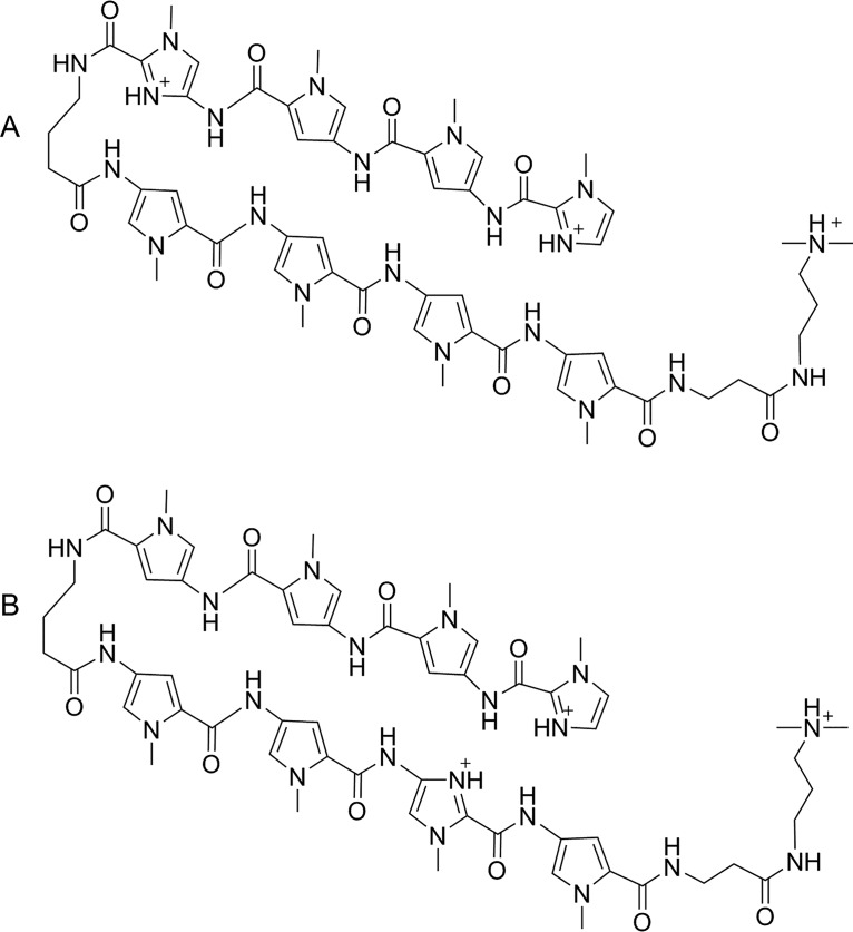
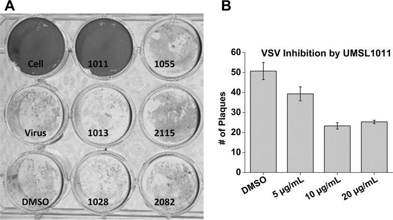
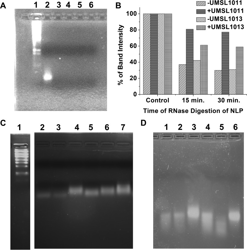
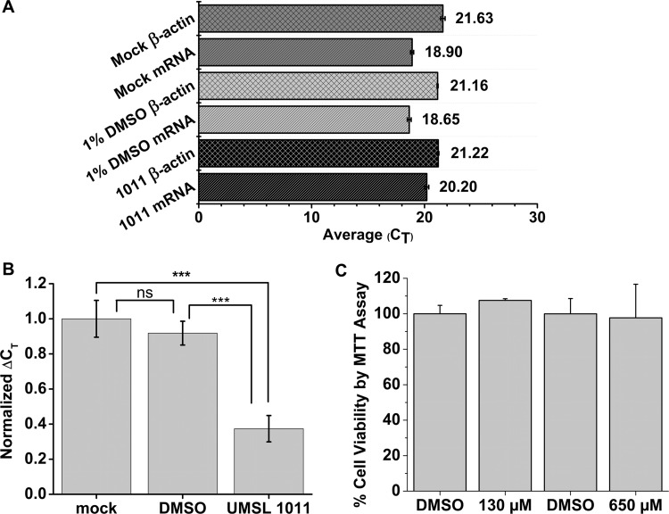
Polyamides have been shown to bind double-stranded DNA by complementing the curvature of the minor groove and forming various hydrogen bonds with DNA. Several polyamide molecules have been found to have potent antiviral activities against papillomavirus, a double-stranded DNA virus. By analogy, we reason that polyamides may also interact with the structured RNA bound in the nucleocapsid of a negative-strand RNA virus. Vesicular stomatitis virus (VSV) was selected as a prototype virus to test this possibility since its genomic RNA encapsidated in the nucleocapsid forms a structure resembling one strand of an A-form RNA duplex. One polyamide molecule, UMSL1011, was found to inhibit infection of VSV. To confirm that the polyamide targeted the nucleocapsid, a nucleocapsid-like particle (NLP) was incubated with UMSL1011. The encapsidated RNA in the polyamide-treated NLP was protected from thermo-release and digestion by RNase A. UMSL1011 also inhibits viral RNA synthesis in the intracellular activity assay for the viral RNA-dependent RNA polymerase. The crystal structure revealed that UMSL1011 binds the structured RNA in the nucleocapsid. The conclusion of our studies is that the RNA in the nucleocapsid is a viable antiviral target of polyamides. Since the RNA structure in the nucleocapsid is similar in all negative-strand RNA viruses, polyamides may be optimized to target the specific RNA genome of a negative-strand RNA virus, such as respiratory syncytial virus and Ebola virus.IMPORTANCE Negative-strand RNA viruses (NSVs) include several life-threatening pathogens, such as rabies virus, respiratory syncytial virus, and Ebola virus. There are no effective antiviral drugs against these viruses. Polyamides offer an exceptional opportunity because they may be optimized to target each NSV. Our studies on vesicular stomatitis virus, an NSV, demonstrated that a polyamide molecule could specifically target the viral RNA in the nucleocapsid and inhibit viral growth. The target specificity of the polyamide molecule was proved by its inhibition of thermo-release and RNA nuclease digestion of the RNA bound in a model nucleocapsid, and a crystal structure of the polyamide inside the nucleocapsid. This encouraging observation provided the proof-of-concept rationale for designing polyamides as antiviral drugs against NSVs.

Keywords: RNA structure; antiviral agents; negative-strand RNA virus; nucleocapsid; viral RNA synthesis.


Figures

FIG 1
FIG 1
RNA structure in VSV NLP. (A) Ribbon and stick drawings of VSV N in complex with RNA. The N-terminal lobe is shown as ribbons in green, and the C-terminal lobe is indicated in yellow. The N-terminal arm and the C-terminal loop elements are labeled. The RNA structure in the center is shown as a stick model. (B) Cartoon diagram of a single subunit in the VSV NLP from the perspective of the N-lobe. This shows the RNA base stacking that occurs within the NLP. All drawings were created with PyMOL (51).
FIG 2
FIG 2
(A) Cation of UMSL1011 isolated as a tris(TFA) salt. (B) Cation of UMSL1013 isolated as a tris(TFA) salt.
FIG 3
FIG 3
VSV plaque assays. (A) An initial screen of six polyamide compounds (20 μg/ml) was performed. Controls of uninfected cells, medium only, and 1% DMSO were included. The compound names were labeled for each well. (B) After virus adsorption for 1 h, UMSL1011 at different concentrations was added to the agarose overlap. DMSO was used as the solvent negative control. Experiments were performed in triplicate, and the plaques were counted at 18 h postinfection.
FIG 4
FIG 4
Melting curve analyses. VSV NLP was incubated with 200 μg/ml (1.3 mM) UMSL1011 (right column), with 200 μg/ml UMSL1013 (middle column), and in 1% DMSO (left column). NLP was at a concentration of 1.2 mg/ml in 50 mM Tris–300 mM NaCl (pH 7.5). Both SYBR Safe and SYPRO Orange were added to the NLP sample (light gray and dark gray traces, respectively). The temperature was scanned at 0.025°C/s using a QuantStudio 3 instrument. The first- and second-derivative graphs are presented. Tfree (68.9°C; equivalent to Tm) was determined from the second-derivative graph. The left column shows the results from the same experiment and presentation as the right column, except that the NLP was incubated with 1% DMSO. Tfree (66.8°C) was determined from the second-derivative graph. To obtain a negative control, the Tfree measurements were repeated with an inactive compound, UMSL1013 (central column). The Tfree value was essentially the same as that for the DMSO control. All errors are shown as standard deviations.
FIG 5
FIG 5
RNA protection study. (A) RNA freed from NLP (Lane 2) was incubated with 15 μg/ml RNase A with (lanes 3 and 4) and without (lanes 5 and 6) 200 μg/ml (1.3 mM) UMSL1011 at 22°C for 30 min (lanes 2 and 4) and 60 min (lanes 3 and 5), respectively. RNA markers (lane 1) are included. (B) Bar graph quantifying the drop in intensity of each band in the gels shown in panels in C and D. Both + and −UMSL1011 and + and −UMSL1013 data are shown compared to each undigested control, which is set at 100%. The program GelAnalyzer 2010a was used to quantify the brightness and overall peak area of each gel, which were normalized on a percentage based on the control. (C) NLP was incubated with 1 mg/ml RNase A at 42°C without (lanes 2 to 4) or with (lanes 5 to 7) 200 μg/ml (1.3 mM) UMSL1011. Lanes 2 and 5, 30-min digestion; lanes 3 and 6, 15-min digestion. Lanes 4 and 7 had no RNase A. Lane 1, RNA markers (200 bases as the lowest band versus 90 bases of RNA from the NLP). (D) Control study with an inactive compound. UMSL1013 (lanes 4 to 6) at 200 μg/ml was incubated with NLP (lane 4, 15 min; lane 5, 30 min). Lane 6 has no RNase A. Lanes 1 to 3 correspond to the same reactions in the presence of 1% DMSO.
FIG 6
FIG 6
Intracellular vRdRp assays and cytotoxicity study. (A) Raw CT values of mRNA transcripts from the minigenome assay and reporter gene, β-actin. Each CT value is taken as an average of three qPCRs, and error bars represent the standard deviations. (B) Normalized ΔCT values to the Mock, set as 1. Each ΔCT was calculated by subtracting the experimental CT value to its subsequent β-actin CT, and the error bars represent the standard deviations. This shows a significant decrease in mRNA transcripts when UMSL1011 is added. ***, P < 0.001; ns (not significant), P > 0.05. (C) Cytotoxicity studies. Monolayers of HeLa cells were incubated with 130 and 650 μM UMSL1011, respectively, for 25 h. We used 1% DMSO as the solvent control. Cell viability was measured using MTT assays. Experiments were carried out in triplicate, and error bars represent the standard deviations.
FIG 7
FIG 7
Crystal structure of UMSL1011 bound to the NLP. (A) 2mFo-dFc map (shown in blue) at 1 sigma of UMSL1011 (shown in green) bound to the NLP. The magenta sticks are nearby amino acids of the NLP, while the RNA is modeled in cyan. The transparent surface reconstruction shows the pocket for which UMSL1011 binds. (B) Hydrogen bonds made by UMSL1011 (shown in green) to both the encapsidated RNA (cyan) and the protein (magenta). (C) Electrostatic surface potential of UMSL1011 calculated by APBS and visualized in Chimera (52, 53). The colored bar below shows the relative charge, with red indicating electron dense and blue indicating electron deficient. (D) Two-dimensional cartoon outlining the hydrogen bonds between UMSL1011 and the RNA[poly(rU)]/protein. Protein contacts are shown with the Gln318 amide carbonyl backbone and the Nε of Arg312.
FIG 8
FIG 8
(A) Overlay of 2GIC and UMSL1011 bound crystal structure. UMSL1011 is indicated in green, while, as described above, magenta and cyan represent the protein and RNA, respectively. The 2GIC structure is yellow. There are few to no structural differences in the local area where UMSL1011 binds. (B) Reconstruction of the NLP ring. On the left-hand side is 2GIC (in yellow), and on the right-hand side is UMSL1011 structure (in cyan). An overall global tightening of the ring by 2.3 Å is observed when measuring the inner diameter from E-chain to E-chain, corresponding to the largest change in the ring diameter.

References

    1. Bont L, Checchia PA, Fauroux B, Figueras-Aloy J, Manzoni P, Paes B, Simoes EA, Carbonell-Estrany X. 2016. Defining the epidemiology and burden of severe respiratory syncytial virus infection among infants and children in western countries. Infect Dis Ther 5:271–298. doi:10.1007/s40121-016-0123-0. - DOI - PMC - PubMed
    1. Appolinario CM, Jackson AC. 2015. Antiviral therapy for human rabies. Antiviral Ther 20:1–10. doi:10.3851/IMP2851. - DOI - PubMed
    1. Balmith M, Faya M, Soliman ME. 2017. Ebola virus: a gap in drug design and discovery—an experimental and computational perspective. Chem Biol Drug Design 89:297–308. doi:10.1111/cbdd.12870. - DOI - PubMed
    1. Wu W, Liu S. 2017. The drug targets and antiviral molecules for treatment of Ebola virus infection. Curr Top Med Chem 17:361–370. doi:10.2174/1568026616666160829161318. - DOI - PubMed
    1. Green TJ, Zhang X, Wertz GW, Luo M. 2006. Structure of the vesicular stomatitis virus nucleoprotein-RNA complex. Science 313:357–360. doi:10.1126/science.1126953. - DOI - PubMed

Publication types

LinkOut - more resources