. 2018 Mar 28;92(8):e02211-17.
doi: 10.1128/JVI.02211-17. Print 2018 Apr 15.

N-Terminomics TAILS Identifies Host Cell Substrates of Poliovirus and Coxsackievirus B3 3C Proteinases That Modulate Virus Infection

Affiliations

N-Terminomics TAILS Identifies Host Cell Substrates of Poliovirus and Coxsackievirus B3 3C Proteinases That Modulate Virus Infection

Julienne M Jagdeo et al. J Virol. .

Abstract

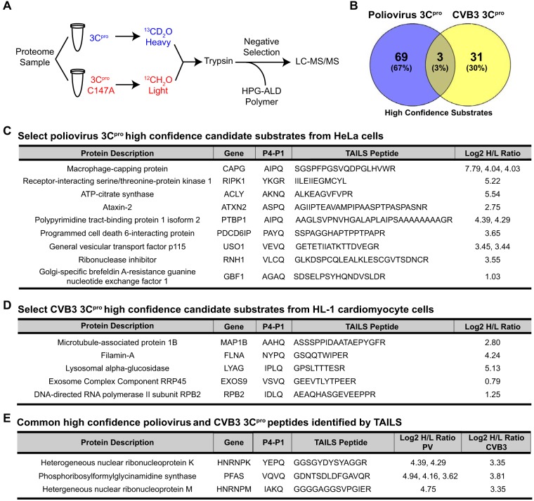
Enteroviruses encode proteinases that are essential for processing of the translated viral polyprotein. In addition, viral proteinases also target host proteins to manipulate cellular processes and evade innate antiviral responses to promote replication and infection. Although some host protein substrates of enterovirus proteinases have been identified, the full repertoire of targets remains unknown. We used a novel quantitative in vitro proteomics-based approach, termed terminal amine isotopic labeling of substrates (TAILS), to identify with high confidence 72 and 34 new host protein targets of poliovirus and coxsackievirus B3 (CVB3) 3C proteinases (3Cpros) in HeLa cell and cardiomyocyte HL-1 cell lysates, respectively. We validated a subset of candidate substrates that are targets of poliovirus 3Cproin vitro including three common protein targets, phosphoribosylformylglycinamidine synthetase (PFAS), hnRNP K, and hnRNP M, of both proteinases. 3Cpro-targeted substrates were also cleaved in virus-infected cells but not noncleavable mutant proteins designed from the TAILS-identified cleavage sites. Knockdown of TAILS-identified target proteins modulated infection both negatively and positively, suggesting that cleavage by 3Cpro promotes infection. Indeed, expression of a cleavage-resistant mutant form of the endoplasmic reticulum (ER)-Golgi vesicle-tethering protein p115 decreased viral replication and yield. As the first comprehensive study to identify and validate functional enterovirus 3Cpro substrates in vivo, we conclude that N-terminomics by TAILS is an effective strategy to identify host targets of viral proteinases in a nonbiased manner.IMPORTANCE Enteroviruses are positive-strand RNA viruses that encode proteases that cleave the viral polyprotein into the individual mature viral proteins. In addition, viral proteases target host proteins in order to modulate cellular pathways and block antiviral responses in order to facilitate virus infection. Although several host protein targets have been identified, the entire list of proteins that are targeted is not known. In this study, we used a novel unbiased proteomics approach to identify ∼100 novel host targets of the enterovirus 3C protease, thus providing further insights into the network of cellular pathways that are modulated to promote virus infection.

Keywords: RNA replication; coxsackievirus; enterovirus; plus-strand RNA virus; poliovirus; proteases; proteinase; proteomics.


Figures

FIG 1
FIG 1
High-confidence candidate substrates of poliovirus and CVB3 3Cpro identified by TAILS. (A) TAILS 3Cpro workflow. Proteome sample (500 μg) extracted from HeLa or HL-1 cell extracts was incubated with poliovirus 3Cpro or CVB3 3Cpro (100 ng/μl), respectively, followed by isotopic dimethylation labeling and TAILS. High-confidence substrates were determined by box plot-and-whiskers analysis of the quantified heavy/light (H/L) ratio of dimethylation-labeled semitryptic neo-N terminus peptides. HPG-ALD, hyperbranched polyglycerol-aldehyde. (B) Venn diagram illustrating the percentage of common high-confidence substrates identified between TAILS analysis of poliovirus and CVB3 3Cpro. (C and D) Select high-confidence substrates of poliovirus 3Cpro from HeLa cell extracts and of CVB3 3Cpro from HL-1 cardiomyocytes. (E) Common peptides identified among both the poliovirus and CVB3 3Cpro list of high-confidence substrates. Peptides listed are statistically significant high-H/L semitryptic neo-N terminus peptides. All high-confidence substrates are listed in Tables S5 and S9 in the supplemental material.
FIG 2
FIG 2
In vitro cleavage assay of known enterovirus 3Cpro substrates. Immunoblot showing cleavage of G3BP1 (top left) and PABP (bottom) following an in vitro cleavage assay with HeLa cell lysates incubated with wild-type (WT) or C147A mutant poliovirus (PV) 3Cpro at the times indicated. Immunoblotting was performed following cleavage of TDP-43 (top right) in HL-1 cell lysates incubated with wild-type or C147A mutant coxsackievirus (CVB3) 3Cpro. cp, cleavage product.
FIG 3
FIG 3
Consensus cleavage site analysis of poliovirus and CVB3 3Cpro high-confidence substrate peptides. Sequence logos of the 10 amino acids positioned directly upstream (P1 to P10) and downstream (P1′ to P10′) of the TAILS-identified peptides for poliovirus 3Cpro (A) and CVB3 3Cpro (B).
FIG 4
FIG 4
Validation of TAILS high-confidence substrates by in vitro cleavage assay. (Left) HeLa cell lysates were incubated with purified wild-type or mutant (C147A) poliovirus 3Cpro (100 ng/μl). Proteins were loaded on an SDS-PAGE gel, and cleavage was assessed by immunoblotting. (Right) Schematics of corresponding high-confidence candidate substrates, indicating key domains, the position of TAILS-predicted cleavage sites, and the four amino acid positions directly upstream (P1 to P4) and downstream (P1′ to P4′) of the cleavage site. The predicted molecular masses of the cleavage protein fragments are shown below. cp, cleavage product; N, N-terminal cleavage product; C, C-terminal cleavage product; GAT, glutamine amidotransferase; KH, hnRNP K homology; PRD, proline rich domain; GTC, Golgi tethering complex; HUS, homology upstream of Sec7; HDS, homology downstream of Sec7; RHIM, RIP homotypic interaction motif; DCB, dimerization and cyclophilin binding; α-TUB, α-tubulin; Ab, antibody.
FIG 5
FIG 5
Validation of TAILS-predicted cleavage site by in vitro cleavage assay. (A) Schematic of a cytomegalovirus (CMV) promoter-driven mammalian expression construct containing 3×FLAG and 3×HA fused in-frame with the full-length candidate substrate. (B) Lysates from HeLa cells expressing the FLAG-HA-tagged wild-type or mutant candidate substrate were incubated with wild-type or mutant poliovirus 3Cpro and immunoblotted for FLAG. N, N-terminal cleavage product.
FIG 6
FIG 6
Cleavage of candidate substrates under virus infection. (A) HeLa cells were mock or poliovirus infected (MOI of 10) for the indicated times. (B) HL-1 cells were mock or CVB3 infected (MOI of 50) for 12 h. Candidate substrate, viral structural protein VP1, and α-tubulin were assessed by immunoblotting. (C) HeLa cells transfected with wild-type or mutant FLAG-HA constructs of candidate substrates were mock or poliovirus infected (MOI of 10) for 7 h. Lysates were immunoblotted with FLAG. hpi, hours postinfection; cp, cleavage product; N, N-terminal cleavage product; C, C-terminal cleavage product.
FIG 7
FIG 7
Cleavage of candidate substrates in poliovirus-infected HeLa cells in the presence of zVAD-FMK. HeLa cells were infected with poliovirus (MOI of 10) in the presence of DMSO or 50 μM zVAD-FMK (7 hpi). Candidate proteins were detected by immunoblotting using the indicated antibody. N, N-terminal cleavage product; C, C-terminal cleavage product.
FIG 8
FIG 8
Candidate substrates identified by TAILS modulate poliovirus infection. HeLa cells were transfected with either a scrambled (siSCX) or a candidate-specific siRNA (si- prefix) for 24 to 72 h, followed by poliovirus infection (MOI of 0.1) for 7 h. Titers of extracellular and intracellular virus were determined by plaque assay, and titers were calculated as the number of PFU per milliliter from at least three independent experiments. (*, P < 0.05; **, P < 0.01). ND, no statistically significant difference. Western blots of the indicated proteins are shown below.
FIG 9
FIG 9
p115 facilitates poliovirus infection. HeLa cells were transfected with either a scrambled (siSCX) or p115 (si-p115) siRNA for 48 h, followed by poliovirus infection (MOI of 1) for the indicated times. (A) Immunofluorescence of endogenous p115 in poliovirus-infected HeLa cells for the times indicated (left). Cells were permeabilized, fixed, and costained for p115 (green; C-terminal antibody) and DNA (blue; Hoechst). An image of HeLa cells stained for viral RNA using an anti-dsRNA antibody at 5 hpi is shown to demonstrate the efficiency of infection at an MOI of 10 (right). (B) Immunoblots of p115, poliovirus structural protein VP1, and α-tubulin are shown. A representative gel is shown from three independent experiments. (C) Northern blot analysis of poliovirus genomic RNA from infected HeLa cells expressing a scrambled (siSCX) or p115 (si-p115) siRNA. GAPDH, glyceraldehyde-3-phosphate dehydrogenase.
FIG 10
FIG 10
Cleavage of p115 modulates GBF1 association with p115. HeLa cells were transfected with either wild-type (WT) or QG832EP GFP-p115 for 48 h, and expression was assessed by immunoblotting using an anti-p115 antibody in uninfected HeLa cells (A) and following poliovirus infection (MOI of 50) for the indicated times (B). Immunoblots of GFP, VP1, and α-tubulin are shown. untr, untransfected. (C and D) FACS-sorted HeLa cells expressing either wild-type (WT) or QG832EP GFP-p115 were poliovirus infected (MOI of 50) for 8 h. Cells were permeabilized, fixed, and costained with GBF1 (blue) and dsRNA (red) antibodies. Representative images from at least two independent experiments are shown for GBF1 and GFP-p115 (C) and dsRNA and GFP-p115 (D). Dot plot graphs show the Manders' correlation coefficient calculated for the fraction of GBF1 overlapping GFP-p115 (C) and the fraction of GFP-p115 overlapping dsRNA (D) in cells expressing wild-type GFP-p115 (n = 46) and QG832EP GFP-p115 (n = 25). Shown are quantitations of a representative experiment that showed a reproducible trend from three independent experiments. *, P < 0.05; **, P < 0.01.
FIG 11
FIG 11
Subcellular localization of wild-type and QG832EP GFP-p115 in HeLa cells. HeLa cells were transfected with either wild-type or QG832EP GFP-p115 for 48 h. Cells were then fixed, permeabilized, and stained for Golgin-97 (red) or GM130 (red), and DNA (blue); 4′,6′-diamidino-2-phenylindole [DAPI].
FIG 12
FIG 12
Subcellular localization of wild-type and QG832EP GFP-p115 and GBF1 in HeLa cells during poliovirus infection. (A) FACS-sorted HeLa cells expressing either wild-type or QG832EP GFP-p115 were mock or PV infected (MOI of 50) for 8 h. Cells were permeabilized, fixed, and costained with GBF1 (blue) and dsRNA (red) antibodies. (B) Representative images from at least two independent experiments are shown for the merged images of GBF1 and dsRNA. (C) Dot plot graphs of the Manders' correlation coefficient calculated for the fraction of GBF1 overlapping dsRNA. Shown are quantitations of a representative experiment that showed a reproducible trend from three independent experiments.
FIG 13
FIG 13
Cleavage of p115 promotes poliovirus infection. (A) Intracellular and extracellular viral titers following poliovirus infection at an MOI of 1 at 8, 12, 16, and 20 hpi of FACS-sorted HeLa cells transfected with wild-type or QG832EP mutant GFP-p115. Average PFU counts ± SD are shown from three independent experiments. **, P < 0.001; *, P < 0.01. (B) Northern blot analysis of poliovirus genomic RNA in FACS-sorted HeLa cells transfected with wild-type or QG832EP mutant GFP-p115 and infected with poliovirus (MOI of 5).

References

    1. Turk B, Turk D, Turk V. 2012. Protease signalling: the cutting edge. EMBO J 31:1630–1643. doi: 10.1038/emboj.2012.42. - DOI - PMC - PubMed
    1. Lin JY, Chen TC, Weng KF, Chang SC, Chen LL, Shih SR. 2009. Viral and host proteins involved in picornavirus life cycle. J Biomed Sci 16:103. doi: 10.1186/1423-0127-16-103. - DOI - PMC - PubMed
    1. Racaniello VR. 2006. One hundred years of poliovirus pathogenesis. Virology 344:9–16. doi: 10.1016/j.virol.2005.09.015. - DOI - PubMed
    1. Marchant D, Si X, Luo H, McManus B, Yang D. 2008. The impact of CVB3 infection on host cell biology. Curr Top Microbiol Immunol 323:177–198. - PubMed
    1. Garmaroudi FS, Marchant D, Hendry R, Luo H, Yang D, Ye X, Shi J, McManus BM. 2015. Coxsackievirus B3 replication and pathogenesis. Future Microbiol 10:629–653. doi: 10.2217/fmb.15.5. - DOI - PubMed

Publication types

MeSH terms

Substances