

Lacunar Infarcts, but Not Perivascular Spaces, Are Predictors of Cognitive Decline in Cerebral Small-Vessel Disease
- PMID: 29438074
- PMCID: PMC5832012
- DOI: 10.1161/STROKEAHA.117.017526
Lacunar Infarcts, but Not Perivascular Spaces, Are Predictors of Cognitive Decline in Cerebral Small-Vessel Disease
Abstract
Background and purpose: Cerebral small-vessel disease is a major cause of cognitive impairment. Perivascular spaces (PvS) occur in small-vessel disease, but their relationship to cognitive impairment remains uncertain. One reason may be difficulty in distinguishing between lacunes and PvS. We determined the relationship between baseline PvS score and PvS volume with change in cognition over a 5-year follow-up. We compared this to the relationship between baseline lacune count and total lacune volume with cognition. In addition, we examined change in PvS volume over time.
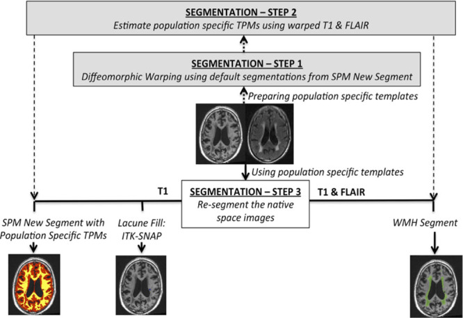
Methods: Data from the prospective SCANS study (St Georges Cognition and Neuroimaging in Stroke) of patients with symptomatic lacunar stroke and confluent leukoaraiosis were used (n=121). Multimodal magnetic resonance imaging was performed annually for 3 years and neuropsychological testing annually for 5 years. Lacunes were manually identified and distinguished from PvS. PvS were rated using a validated visual rating scale, and PvS volumes calculated using T1-weighted images. Linear mixed-effect models were used to determine the impact of PvS and lacunes on cognition.
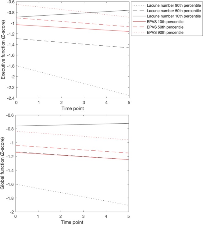
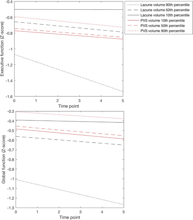
Results: Baseline PvS scores or volumes showed no association with cognitive indices. No change was detectable in PvS volumes over the 3 years. In contrast, baseline lacunes associated with all cognitive indices and predicted cognitive decline over the 5-year follow-up.
Conclusions: Although a feature of small-vessel disease, PvS are not a predictor of cognitive decline, in contrast to lacunes. This study highlights the importance of carefully differentiating between lacunes and PvS in studies investigating vascular cognitive impairment.
Keywords: cerebral small vessel diseases; cognition; leukoaraiosis; magnetic resonance imaging; neuroimaging.
© 2018 The Authors.
Figures
Comment in
-
Letter by Feng et al Regarding Article, "Lacunar Infarcts, but Not Perivascular Spaces, Are Predictors of Cognitive Decline in Cerebral Small-Vessel Disease".Stroke. 2018 Aug;49(8):e276. doi: 10.1161/STROKEAHA.118.021461. Stroke. 2018. PMID: 29967013 No abstract available.
References
-
- Pantoni L. Cerebral small vessel disease: from pathogenesis and clinical characteristics to therapeutic challenges. Lancet Neurol. 2010;9:689–701. doi: 10.1016/S1474-4422(10)70104-6. - PubMed
-
- Wardlaw JM, Smith EE, Biessels GJ, Cordonnier C, Fazekas F, Frayne R, et al. STandards for ReportIng Vascular changes on nEuroimaging (STRIVE v1) Neuroimaging standards for research into small vessel disease and its contribution to ageing and neurodegeneration. Lancet Neurol. 2013;12:822–838. doi: 10.1016/S1474-4422(13)70124-8. - PMC - PubMed
-
- Fergus N, Doubal AMJM. Enlarged perivascular spaces on MRI are a feature of cerebral small vessel disease. Stroke J. Cereb. Circ. 2010;41:450–454. - PubMed
-
- Yao M, Zhu YC, Soumaré A, Dufouil C, Mazoyer B, Tzourio C, et al. Hippocampal perivascular spaces are related to aging and blood pressure but not to cognition. Neurobiol Aging. 2014;35:2118–2125. doi: 10.1016/j.neurobiolaging.2014.03.021. - PubMed
Publication types
MeSH terms
Grants and funding
LinkOut - more resources
Full Text Sources
Other Literature Sources
Medical
Research Materials
