. 2018 Apr 20;29(16):165101.
doi: 10.1088/1361-6528/aaaf16.

Magnetic resonance and photoacoustic imaging of brain tumor mediated by mesenchymal stem cell labeled with multifunctional nanoparticle introduced via carotid artery injection

Affiliations

Magnetic resonance and photoacoustic imaging of brain tumor mediated by mesenchymal stem cell labeled with multifunctional nanoparticle introduced via carotid artery injection

Yang Qiao et al. Nanotechnology. .

Abstract

Objective: To evaluate the feasibility of visualizing bone marrow-derived human mesenchymal stem cells (MSCs) labeled with a gold-coated magnetic resonance (MR)-active multifunctional nanoparticle and injected via the carotid artery for assessing the extent of MSC homing in glioma-bearing mice.

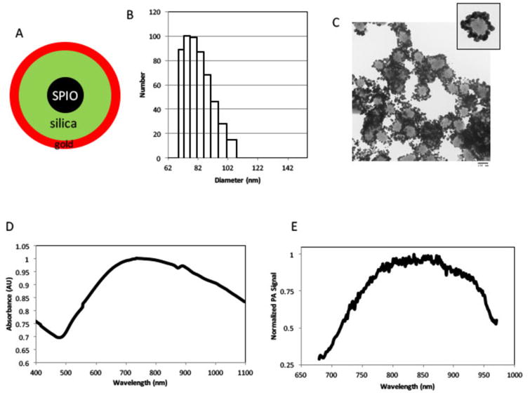
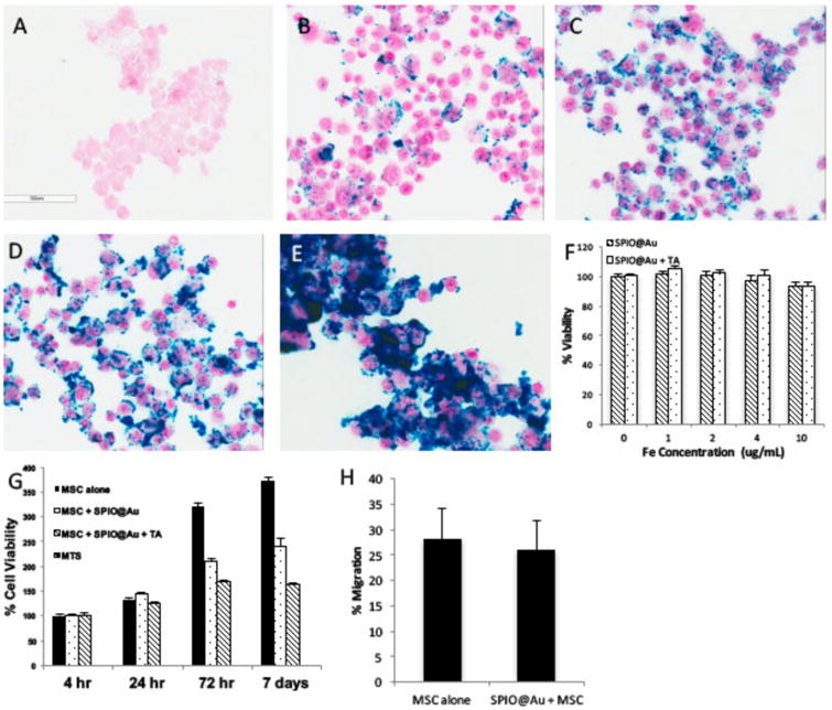
Materials and methods: Nanoparticles containing superparamagnetic iron oxide coated with gold (SPIO@Au) with a diameter of ∼82 nm and maximum absorbance in the near infrared region were synthesized. Bone marrow-derived MSCs conjugated with green fluorescent protein (GFP) were successfully labeled with SPIO@Au at 4 μg ml-1 and injected via the internal carotid artery in six mice bearing orthotopic U87 tumors. Unlabeled MSCs were used as a control. The ability of SPIO@Au-loaded MSCs to be imaged using MR and photoacoustic (PA) imaging at t = 0 h, 2 h, 24 h, and 72 h was assessed using a 7 T Bruker Biospec experimental MR scanner and a Vevo LAZR PA imaging system with a 5 ns laser as the excitation source. Histological analysis of the brain tissue was performed 72 h after MSC injection using GFP fluorescence, Prussian blue staining, and hematoxylin-and-eosin staining.

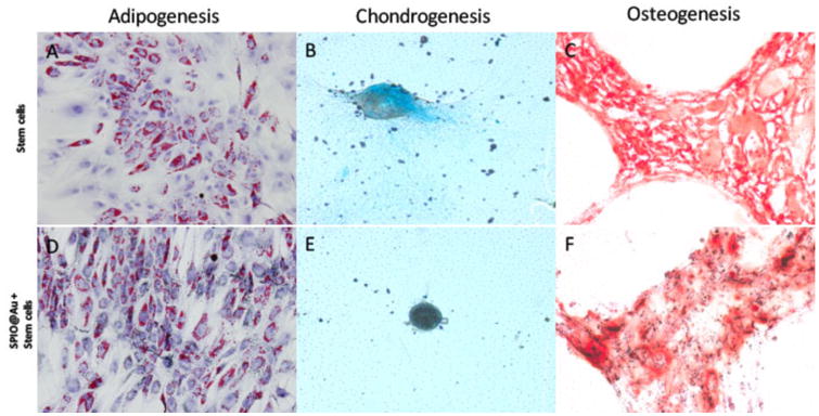
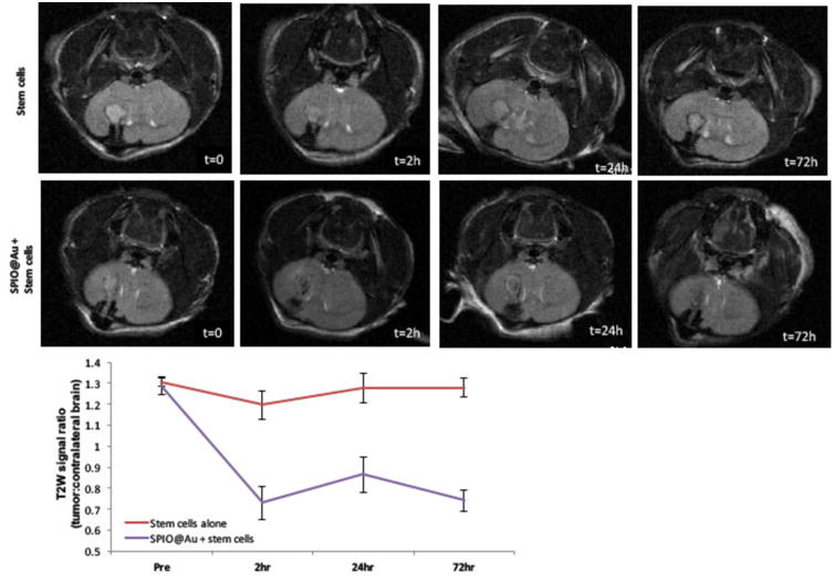
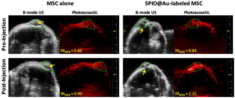
Results: MSCs labeled with SPIO@Au at 4 μg ml-1 did not exhibit cell death or any adverse effects on differentiation or migration. The PA signal in tumors injected with SPIO@Au-loaded MSCs was clearly more enhanced post-injection, as compared with the tumors injected with unlabeled MSCs at t = 72 h. Using the same mice, T2-weighted MR imaging results taken before injection and at t = 2 h, 24 h, and 72 h were consistent with the PA imaging results, showing significant hypointensity of the tumor in the presence of SPIO@Au-loaded MSCs. Histological analysis also showed co-localization of GFP fluorescence and iron, thereby confirming that SPIO@Au-labeled MSCs continue to carry their nanoparticle payloads even at 72 h after injection.

Conclusions: Our results demonstrated the feasibility of tracking carotid artery-injected SPIO@Au-labeled MSCs in vivo via MR and PA imaging.


Figures

Figure 1
Figure 1
Synthesis and characterization of SPIO@Au nanoparticles. (A) Schematic illustration of SPIO@Au nanoparticle containing superparamagnetic iron oxide (SPIO) core, silica, and gold coating. (B) Dynamic light scattering analysis and (C) transmission electron microscopic image of SPIO@Au indicating the size of the nanoparticle. (D) Ultraviolet-visible light absorption spectrum and (E) photoacoustic (PA) profile of SPIO@Au.
Figure 2
Figure 2
In vitro characterization of SPIO@Au-labeled MSCs. (A-E) Bright-field micrographs of Prussian blue-stained MSCs loaded with different concentrations of SPIO@Au (A, no SPIO@Au; B, 1 μg/mL; C, 2 μg/mL; D, 4 μg/mL; E, 10 μg/mL + 0.1% lipofectamine). (F) Quantification of the viability of MSCs loaded with different concentrations of SPIO@Au with and without a transfecting agent (TA) at 4 h. Data are shown as means ± standard deviations (error bars). (G) Cell viability at 4 μg/mL SPIO@Au with or without 0.1% TA at different time points after MSC loading (t = 4 h, 24 h, 72 h, and 7 days). (H) Percent migration of labeled versus unlabeled MSCs (p = 0.16).
Figure 3
Figure 3
Micrographs comparing the ability of MSCs with and without SPIO@Au labeling to differentiate toward adipogenic (A, D), chondrogenic (B, E), and osteogenic (C, F) lineages.
Figure 4
Figure 4
Representative T2-weighted MR images of the mouse brain at various times (t = 0, 2h, 24h, and 72 h) after intra-carotid artery injection of SPIO@Au-loaded MSCs or unlabeled MSCs (top). Quantification of the T2-weighted signal of the tumor against that of the contralateral brain (bottom).
Figure 5
Figure 5
In vivo photoacoustic (PA) imaging of mouse brain at 72 h after intra-carotid artery injection of SPIO@Au-labeled MSCs or MSCs alone. Yellow arrows on B-mode ultrasound (US) images of the brain indicate the bolt placement where the U87 cells were implanted into the brain. The PA images were taken at 810 nm, and the signal intensity was calculated within the green regions of interest. Average PA (PAAVE) values were obtained by averaging all PA intensity values above the signal-noise threshold within the regions of interest.
Figure 6
Figure 6
Histologic analysis after intra-carotid artery injection showing SPIO@Au-loaded MSCs homing to the U87 brain tumor. Hematoxylin and eosin (H&E) staining of the mouse brain with U87 tumor (left). GFP fluorescence microscopy image at 20× magnification showing presence of MSCs (middle). Prussian blue staining of SPIO@Au indicating co-localization of the GFP-labeled MSCs and the iron of the SPIO@Au nanoparticle (right). This co-localization was not observed with the control (GFP-labeled MSCs not loaded with SPIO@Au).

References

    1. Binello E, Germano IM. Stem cells as therapeutic vehicles for the treatment of high-grade gliomas. Neuro Oncol. 2012;14:256–65. - PMC - PubMed
    1. Hamada H, Kobune M, Nakamura K, Kawano Y, Kato K, Honmou O, Houkin K, Matsunaga T, Niitsu Y. Mesenchymal stem cells (MSC) as therapeutic cytoreagents for gene therapy. Cancer Sci. 2005;96:149–56. - PMC - PubMed
    1. Kosztowski T, Zaidi HA, Quiñones-Hinojosa A. Applications of neural and mesenchymal stem cells in the treatment of gliomas. Expert Rev Anticancer Ther. 2009;9:597–612. - PMC - PubMed
    1. Miletic H, et al. Bystander killing of malignant glioma by bone marrow-derived tumor-infiltrating progenitor cells expressing a suicide gene. Mol Ther. 2007;15:1373–81. - PubMed
    1. Nakamizo A, et al. Human bone marrow-derived mesenchymal stem cells in the treatment of gliomas. Cancer Res. 2005;65:3307–18. - PubMed

MeSH terms