Review
. 2018 Feb 13;6(1):16.
doi: 10.3390/diseases6010016.

Diaphragmatic Palsy

Affiliations
Review

Diaphragmatic Palsy

Laxmi Kokatnur et al. Diseases. .

Abstract

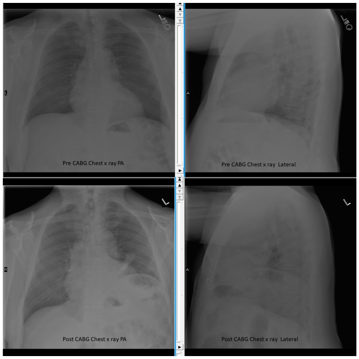
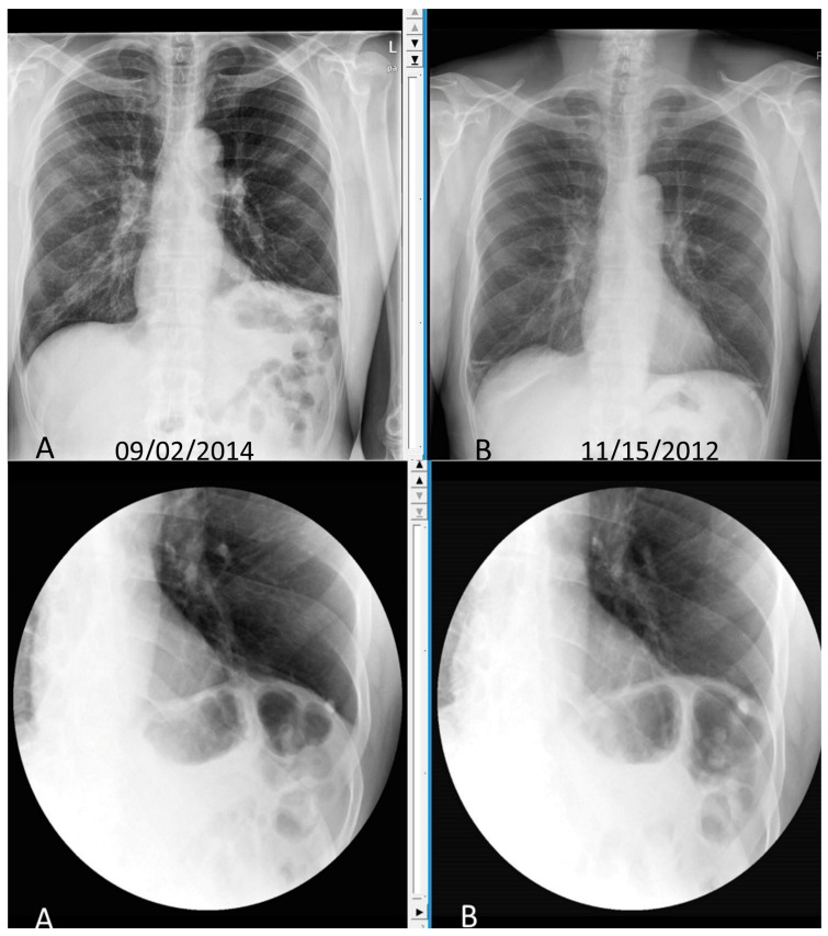
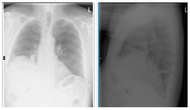
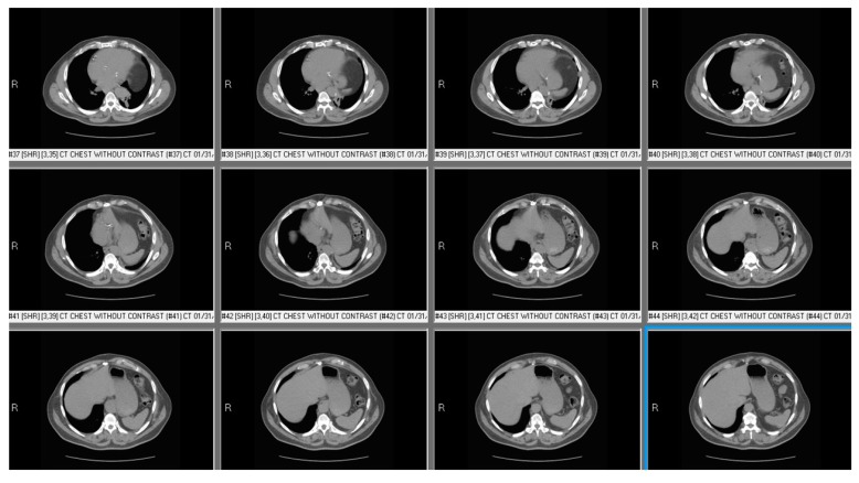
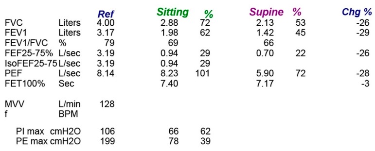
The diaphragm is the primary muscle of respiration, and its weakness can lead to respiratory failure. Diaphragmatic palsy can be caused by various causes. Injury to the phrenic nerve during thoracic surgeries is the most common cause for diaphragmatic palsy. Depending on the cause, the symptoms of diaphragmatic palsies vary from completely asymptomatic to disabling dyspnea requiring mechanical ventilation. On pulmonary function tests, there will be a decrease in the maximum respiratory muscle power. Spirometry shows reduced lung functions and a significant drop of lung function in supine position is typical of diaphragmatic palsy. Diaphragmatic movements with respiration can be directly visualized by fluoroscopic examination. Currently, this test is being replaced by bedside thoracic ultrasound examination, looking at the diaphragmic excursion with deep breathing or sniffing. This test is found to be equally efficient, and without risks of ionizing radiation of fluoroscope. Treatment of diaphragmatic palsy depends on the cause. Surgical approach of repair of diaphragm or nonsurgical approach of noninvasive ventilation has been tried with good success. Overall prognosis of diaphragmatic palsy is good, except when it is related to neuromuscular degeneration conditions.

Keywords: diaphragmatic palsy; phrenic nerve injury; pulmonary function tests; respiratory failure.


Conflict of interest statement

The authors declare no conflict of interest.

Figures

Figure 1
Figure 1
Chest X-ray Pre and Post Cardiac Bypass Surgery (CABG) showing elevated left diaphragm after surgery. The sternal metal suture wires also seen.
Figure 2
Figure 2
Chest X-ray and Static Sniff test of patient with cervical spondylosis leading to phrenic nerve compression and left diaphragmatic palsy. Top panel showing chest X-ray with elevated left diaphragm (A) compared to normal position on chest X-ray done 2 years back (B). Lower panel. Static Sniff test images in expiration (A) and after sniffing (B) showing no movement of diaphragm.
Figure 3
Figure 3
Chest X-ray Posterior-Anterior and Lateral view showing right diaphragm located more than 2 intercostal spaces compared to left side.
Figure 4
Figure 4
Computed Tomography (CT) chest of patient with left diaphragmatic palsy showing abdominal contents beside heart and at higher level compared to liver suggesting left diaphragmatic palsy.
Figure 5
Figure 5
Spirometry of patient with bilateral diaphragmatic palsy showing significant drop of lung functions on supine position.

References

    1. Bordoni B., Zanier E. Anatomic connections of the diaphragm: Influence of respiration on the body system. J. Multidiscip. Healthc. 2013;6:281. doi: 10.2147/JMDH.S45443. - DOI - PMC - PubMed
    1. Schumpelick V., Steinau G., Schlüper I., Prescher A. Surgical embryology and anatomy of the diaphragm with surgical applications. Surg. Clin. 2000;80:213–239. doi: 10.1016/S0039-6109(05)70403-5. - DOI - PubMed
    1. Maish M.S. The diaphragm. Surg. Clin. N. Am. 2010;90:955–968. doi: 10.1016/j.suc.2010.07.005. - DOI - PubMed
    1. Dubé B.P., Dres M. Diaphragm Dysfunction: Diagnostic Approaches and Management Strategies. J. Clin. Med. 2016;5:113. doi: 10.3390/jcm5120113. - DOI - PMC - PubMed
    1. Aguirre V.J., Sinha P., Zimmet A., Lee G.A., Kwa L., Rosenfeldt F. Phrenic nerve injury during cardiac surgery: Mechanisms, management and prevention. Heart Lung Circ. 2013;22:895–902. doi: 10.1016/j.hlc.2013.06.010. - DOI - PubMed

LinkOut - more resources