. 2018 Feb 13;19(2):565.
doi: 10.3390/ijms19020565.

Sjögren-Like Lacrimal Keratoconjunctivitis in Germ-Free Mice

Affiliations

Sjögren-Like Lacrimal Keratoconjunctivitis in Germ-Free Mice

Changjun Wang et al. Int J Mol Sci. .

Abstract

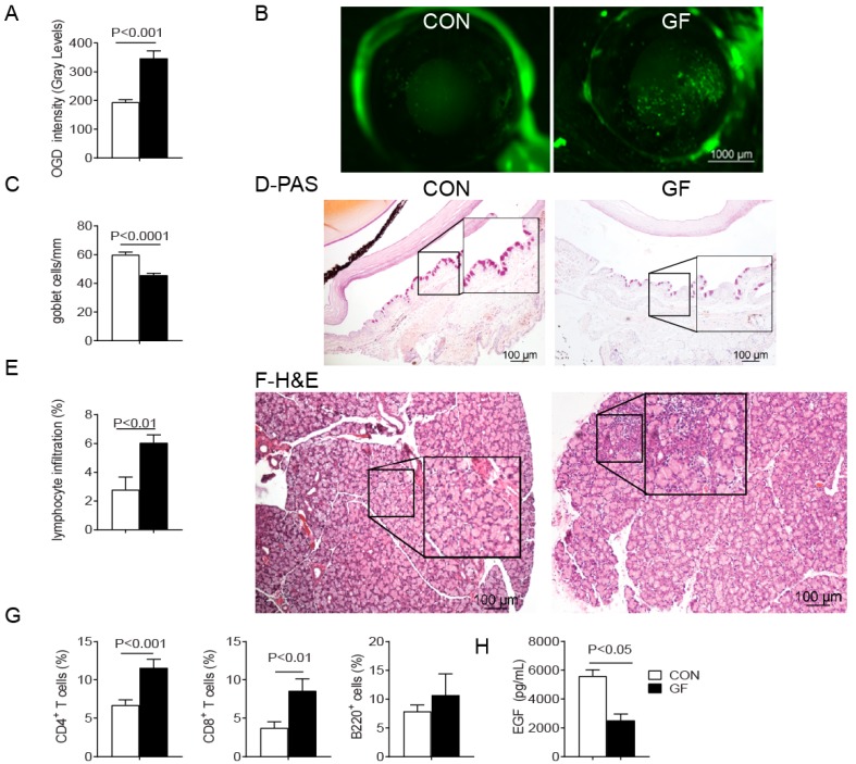
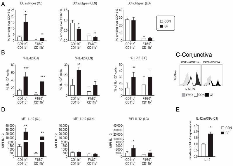
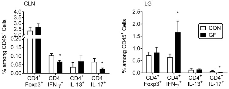
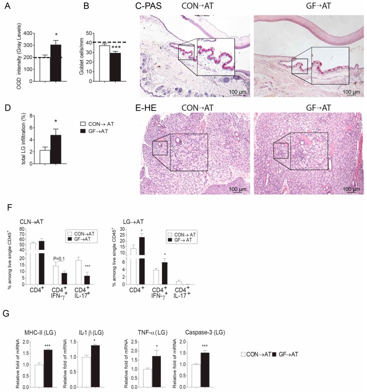
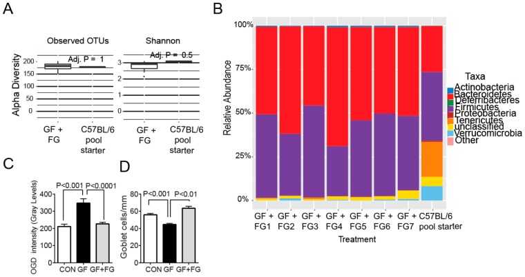
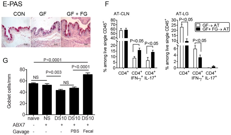
Commensal bacteria play an important role in the formation of the immune system but their role in the maintenance of immune homeostasis at the ocular surface and lacrimal gland remains poorly understood. This study investigated the eye and lacrimal gland phenotype in germ-free and conventional C57BL/6J mice. Our results showed that germ-free mice had significantly greater corneal barrier disruption, greater goblet cell loss, and greater total inflammatory cell and CD4⁺ T cell infiltration within the lacrimal gland compared to the conventionally housed group. A greater frequency of CD4⁺IFN-γ⁺ cells was observed in germ-free lacrimal glands. Females exhibited a more severe phenotype compared to males. Adoptive transfer of CD4⁺ T cells isolated from female germ-free mice into RAG1KO mice transferred Sjögren-like lacrimal keratoconjunctivitis. Fecal microbiota transplant from conventional mice reverted dry eye phenotype in germ-free mice and decreased CD4⁺IFN-γ⁺ cells to levels similar to conventional C57BL/6J mice. These findings indicate that germ-free mice have a spontaneous lacrimal keratoconjunctivitis similar to that observed in Sjögren syndrome patients and demonstrate that commensal bacteria function in maintaining immune homeostasis on the ocular surface. Thus, manipulation of intestinal commensal bacteria has the potential to become a novel therapeutic approach to treat Sjögren Syndrome.

Keywords: Sjögren syndrome; commensal bacteria; dry eye; fecal transplant; germ-free mice; goblet cell.


Conflict of interest statement

All authors state that they have no conflicts of interest.

Figures

Figure 1
Figure 1
Sjögren-like Lacrimal Keratoconjunctivitis on germ-free mice. Conventionally housed mice with complex microbiota served as control (CON) and were compared to germ-free (GF) mice at 8 weeks of age. Both sexes were pooled. (A) Corneal Oregon-Green dextran fluorescence intensity score. Bar graphs show means ± SEM of two independent experiments with four to five animals per experiment (final n = 8–10 animals, mixed sex); (B) Representative images of corneas stained with Oregon Green dextran (OGD); (C) Number of Periodic acid–Schiff (PAS)+ conjunctival goblet cells counted in paraffin-embedded sections expressed as number per millimeter. Bar graph show means ± SEM of two independent experiments with three animals per group, yielding a final sample of six right eyes for each group); (D) Representative images of conjunctiva sections stained with PAS used to generate the bar graph in (C); (E) Total lacrimal gland infiltration measured in haematoxylin and eosin (H&E) stained sections as shown in (F) (n = 6 right lacrimal gland); (F) Representative images of H&E-stained sections of lacrimal gland. Black rectangular insets are a high magnification of dotted square; (G) Flow cytometric analysis of CD4+ and CD8+ T cells and B220+ B cells in lacrimal gland. Right and left extraorbital lacrimal glands from one mouse per group were excised and pooled into a single tube, yielding a final sample of 12 individual lacrimal gland samples divided into two independent experiments with six samples per experiment. Bar graphs show means ± SEM; (H) Tear epidermal growth factor (EGF) concentrations were measured by enzyme-linked immunosorbent assay. Tear washings from both right and left eyes from one mouse per group were collected and pooled into a single tube, yielding a final sample of 12 individual samples per group and divided into three independent experiments with four samples per experiment) Mann-Whitney U test was used for germ-free vs. conventional mice comparisons.
Figure 2
Figure 2
DC frequency and production of IL-12 is altered in germ-free mice. Single cell suspensions of the conjunctiva (CJ), cervical lymph nodes (CLN) and lacrimal glands (LG) were prepared and stained for antigen-presenting cell and macrophage markers followed by intracellular staining for IL-12. Bar graphs are shown as means ± SEM of a representative experiment with 4–5 samples per group. The experiment was repeated once with similar results. (A) Accumulative data showing frequency of CD11b+CD11c+ and F4/80+CD11b+ cells among alive CD45+ gated cells; (B) Accumulative data showing frequency of IL-12+ among viable CD45+ gated cells; (C) Representative overlaid histogram of IL-12 staining in conjunctival CD11b+CD11c+ and F4/80+CD11b+ cells; (D) Accumulative data showing median fluorescence intensity (MIF) of IL-12 in positive cells; (E) Relative fold expression changes of IL-12 in full-thickness conjunctival biopsies. Bar graphs show means ± SD of five samples per group, biological replicates from two independent experiments were averaged. FMO = fluorescence minus one; GF = germ-free; CON = conventional mice; * p < 0.05; ** p < 0.01; *** p < 0.001. Mann–Whitney U test was used for GF vs. CON comparison test.
Figure 3
Figure 3
Increased percentage of Th1+ cells in lacrimal gland of germ-free mice. Percentage of CD4+Foxp3+, Th1 (CD4+IFN-γ+), Th2 (CD4+IL-13+), Th17 (CD4+IL-17+) cells in freshly isolated cervical lymph nodes (CLN) and lacrimal glands (LG) by flow cytometry. Each experiment group consisted of three-to-four samples; showing mean ± SEM of two independent experiments (final n = seven to eight per group). CLN of the same animal were pooled; right and left extraorbital LGs of the same animal were pooled. * p < 0.05 conventional vs. germ-free comparison using Mann-Whitney U test.
Figure 4
Figure 4
CD4+T Cells from germ-free mice transfer Sjögren-like lacrimal Keratoconjunctivitis to immunodeficient RAG1KO mice. CD4+T cells were isolated from spleens and cervical lymph nodes (CLN) from conventionally housed (CON) and germ-free (GF) mice and adoptively transferred (→AT) into RAG1KO mice. Disease severity parameters were evaluated five weeks later. Bar graphs show means ± SEM of a representative experiment with 18–20 samples per group. (A) Corneal barrier function measured by Oregon-Green dextran fluorescence intensity score. Bar graphs show means ± SEM of two independent experiments with four animals per experiment (final n = eight animals, female sex). Dotted line demonstrates OGD fluorescence intensity score in naïve RAG1KO mice; (B) Number of PAS+ conjunctival goblet cells counted in paraffin-embedded sections expressed as number per millimeter. Bar graphs show means ± SEM of two independent experiments with three animals per group, yielding a final sample of six right eyes for each group. Dotted line demonstrates goblet cell density in naïve RAG1KO mice; (C) Representative images of conjunctiva sections stained with PAS (purple cells) used to generate the bar graph in (B). Black rectangular insets are a high magnification of the small demarcated area; (D) Total lacrimal gland infiltration measured in H&E stained sections as shown in (F) (n = six right lacrimal glands); (E) Representative images of haematoxylin and eosin (H&E)-stained sections. Black rectangular insets are a high magnification of small demarcated area; (F) Percentage of total CD4+ T, Th1 (CD4+IFN-γ+), Th2 (CD4+IL-13+), Th17 (CD4+IL-17+) cells in freshly isolated cervical lymph nodes (CLN) and lacrimal glands (LG) by flow cytometry five weeks post-adoptive transfer. Each experiment group consisted of three-to-four samples; showing mean ± SEM of two independent experiments. CLN of the same animal were pooled; right and left extraorbital LGs of the same animal were pooled. Bar graphs show means ± SEM of two independent experiments with four animals per experiment (final n = eight animals, female sex); (G) Relative fold expression changes of MHC II, IL-, TNF-α and Caspase 3 in LG. Bar graphs show means ± SD of six samples per group, biological replicates from two independent experiments were averaged. * p < 0.05; *** p < 0.001Mann-Whitney U GF vs. CON comparison test.
Figure 5
Figure 5
Reconstitution of germ-free mice with commensal bacteria reverses the dry eye phenotype. (AC) Four-week-old female germ-free mice were colonized with a fecal slurry from normal mice (a pool of three mice) by intragastric gavage and sacrificed at eight weeks of age (germ-free + Fecal gavage, GF + FG). (A) Comparison of α diversity between starting inoculum (C57BL/6J) and germ-free conventionalized mice (n = seven) using measures of observed OTUs and Shannon’s diversity index; (B) Relative abundance of major bacterial phyla in the starting inoculum (C57BL/6J pool) used to conventionalize mice and in the gut microbiota from conventionalized germ-free mice that received fecal gavage (n = seven) at eight weeks of age; (C) Corneal Oregon-Green dextran fluorescence intensity score. Bar graphs show means ± SEM of two independent experiments with four animals per experiment (final n = eight animals, female sex). Mann-Whitney U comparison test; (D) Number of PAS+ conjunctival goblet cells counted in paraffin-embedded sections expressed as number per millimeter. Bar graphs show means ± SEM of two independent experiments with three-four animals per group, yielding a final sample of seven right eyes for each group). Mann-Whitney U comparison test; (E) Representative images of conjunctiva sections stained with PAS used to generate the bar graph in (D); (F) Flow cytometry analysis showing the percentage of total CD4+, Th1 (CD4+IFN-γ+), Th17 (CD4+IL-17+) cells in freshly isolated cervical lymph nodes (CLN) and lacrimal glands (LG) in adoptive transfer (→AT) recipients of either germ-free or germ-free + FG CD4+T cells. Each experiment group consisted of three-to-four samples; showing means ± SEM of two independent experiments, yielding a final sample of seven to eight per group. Mann-Whitney U comparison test; (G) Fecal transplant during desiccating stress (DS) rescues goblet cells. Female conventional C57BL/6 mice were left untreated (naïve mice) or received a cocktail of antibiotics (ABX) for seven days. On the morning of the 8th day, mice were switched to normal water and subjected to desiccating stress (DS) for ten days (DS10) and randomized to receive either PBS or oral gavage of fecal material or were left non-stressed. Mice under DS were sacrificed after ten days, and the number of PAS+ cells in the conjunctiva was determined. n = five animals/group. Kruskal-Wallis test followed by Tukey’s post hoc test. NS = non-significant.
Figure 5
Figure 5
Reconstitution of germ-free mice with commensal bacteria reverses the dry eye phenotype. (AC) Four-week-old female germ-free mice were colonized with a fecal slurry from normal mice (a pool of three mice) by intragastric gavage and sacrificed at eight weeks of age (germ-free + Fecal gavage, GF + FG). (A) Comparison of α diversity between starting inoculum (C57BL/6J) and germ-free conventionalized mice (n = seven) using measures of observed OTUs and Shannon’s diversity index; (B) Relative abundance of major bacterial phyla in the starting inoculum (C57BL/6J pool) used to conventionalize mice and in the gut microbiota from conventionalized germ-free mice that received fecal gavage (n = seven) at eight weeks of age; (C) Corneal Oregon-Green dextran fluorescence intensity score. Bar graphs show means ± SEM of two independent experiments with four animals per experiment (final n = eight animals, female sex). Mann-Whitney U comparison test; (D) Number of PAS+ conjunctival goblet cells counted in paraffin-embedded sections expressed as number per millimeter. Bar graphs show means ± SEM of two independent experiments with three-four animals per group, yielding a final sample of seven right eyes for each group). Mann-Whitney U comparison test; (E) Representative images of conjunctiva sections stained with PAS used to generate the bar graph in (D); (F) Flow cytometry analysis showing the percentage of total CD4+, Th1 (CD4+IFN-γ+), Th17 (CD4+IL-17+) cells in freshly isolated cervical lymph nodes (CLN) and lacrimal glands (LG) in adoptive transfer (→AT) recipients of either germ-free or germ-free + FG CD4+T cells. Each experiment group consisted of three-to-four samples; showing means ± SEM of two independent experiments, yielding a final sample of seven to eight per group. Mann-Whitney U comparison test; (G) Fecal transplant during desiccating stress (DS) rescues goblet cells. Female conventional C57BL/6 mice were left untreated (naïve mice) or received a cocktail of antibiotics (ABX) for seven days. On the morning of the 8th day, mice were switched to normal water and subjected to desiccating stress (DS) for ten days (DS10) and randomized to receive either PBS or oral gavage of fecal material or were left non-stressed. Mice under DS were sacrificed after ten days, and the number of PAS+ cells in the conjunctiva was determined. n = five animals/group. Kruskal-Wallis test followed by Tukey’s post hoc test. NS = non-significant.

References

    1. Nolan J. Evaluation of conjunctival and nasal bacterial cultures before intra-ocular operations. Br. J. Ophthalmol. 1967;51:483–485. doi: 10.1136/bjo.51.7.483. - DOI - PMC - PubMed
    1. Fahmy J.A., Moller S., Bentzon M.W. Bacterial flora in relation to cataract extraction. II. Peroperative flora. Acta Ophthalmol. 1975;53:476–494. doi: 10.1111/j.1755-3768.1975.tb01178.x. - DOI - PubMed
    1. Schabereiter-Gurtner C., Maca S., Rolleke S., Nigl K., Lukas J., Hirschl A., Lubitz W., Barisani-Asenbauer T. 16S rDNA-based identification of bacteria from conjunctival swabs by PCR and DGGE fingerprinting. Investig. Ophthalmol. Vis. Sci. 2001;42:1164–1171. - PubMed
    1. Narayanan S., Miller W.L., McDermott A.M. Expression of human β-defensins in conjunctival epithelium: Relevance to dry eye disease. Investig. Ophthalmol. Vis. Sci. 2003;44:3795–3801. doi: 10.1167/iovs.02-1301. - DOI - PMC - PubMed
    1. Zhou L., Huang L.Q., Beuerman R.W., Grigg M.E., Li S.F., Chew F.T., Ang L., Stern M.E., Tan D. Proteomic analysis of human tears: Defensin expression after ocular surface surgery. J. Proteome Res. 2004;3:410–416. doi: 10.1021/pr034065n. - DOI - PubMed

LinkOut - more resources