Review
. 2018 Jul 1;99(1):27-44.
doi: 10.1093/biolre/ioy043.

Estrogen in the male: a historical perspective

Affiliations
Review

Estrogen in the male: a historical perspective

Rex A Hess et al. Biol Reprod. .

Abstract

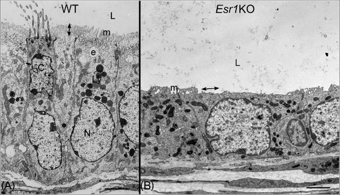
Estrogens have traditionally been considered female hormones. Nevertheless, the presence of estrogen in males has been known for over 90 years. Initial studies suggested that estrogen was deleterious to male reproduction because exogenous treatments induced developmental abnormalities. However, demonstrations of estrogen synthesis in the testis and high concentrations of 17β-estradiol in rete testis fluid suggested that the female hormone might have a function in normal male reproduction. Identification of estrogen receptors and development of biological radioisotope methods to assess estradiol binding revealed that the male reproductive tract expresses estrogen receptor extensively from the neonatal period to adulthood. This indicated a role for estrogens in normal development, especially in efferent ductules, whose epithelium is the first in the male reproductive tract to express estrogen receptor during development and a site of exceedingly high expression. In the 1990s, a paradigm shift occurred in our understanding of estrogen function in the male, ushered in by knockout mouse models where estrogen production or expression of its receptors was not present. These knockout animals revealed that estrogen's main receptor (estrogen receptor 1 [ESR1]) is essential for male fertility and development of efferent ductules, epididymis, and prostate, and that loss of only the membrane fraction of ESR1 was sufficient to induce extensive male reproductive abnormalities and infertility. This review provides perspectives on the major discoveries and developments that led to our current knowledge of estrogen's importance in the male reproductive tract and shaped our evolving concept of estrogen's physiological role in the male.


Figures

Figure 1.
Figure 1.
Immunohistochemical localization of P450 aromatase protein in the mouse testis and epididymis. (A) Aromatase protein was expressed in the cytoplasm of round (RS) and elongated spermatids (ES) in the mouse seminiferous epithelium. (B) Caput epididymal lumen. Aromatase protein was localized in the cytoplasmic droplet (Cd) and along the thin tails of the spermatozoa. Used with Open Access permission from a prior publication [11].
Figure 2.
Figure 2.
Comparison of the effects on testis weight and efferent ductules after treatment with the fungicide carbendazim and in the Esr1 knockout mouse (Esr1KO). Modified with permission from a prior publication [100]. On the left, rat testis weight increases within hours after treatment with carbendazim (red), reaching a peak by day 4 and then decreasing as the testis atrophies. On the right, testis weight increases in Esr1KO mice (red) reaching a peak on day 75, but then decreasing until it has atrophied by day 185. In both cases, testicular atrophy was preceded by increased testis weight due to back-pressure of fluid accumulation caused by disruption of efferent ductule structure and function. (A) Wild-type (WT) efferent ductules showing a normal lumen (L) containing mostly fluid that must be reabsorbed as sperm are transported toward the epididymis. The epithelium (E) has a normal height and is lined by ciliated cells (Ci) with long motile cilia extending into the lumen and by nonciliated cells (N) that have a periodic acid-Schiff's positive border at the lumen where numerous microvilli are present. The connective tissue (C) contains loosely scattered fibroblasts and blood vessels. (B) Efferent ductules from the fungicide carbendazim-treated rat. The lumens (L) are occluded with coagulated sperm and cellular debris. The lining epithelium cannot be distinguished at this magnification due to the number of inflammatory cells (In) surrounding the ductules in the densely populated connective tissue (C), which also appears thickened. In one area, the lining epithelium appears to have begun recanalization (R) or regrowth around the occlusion. (C) Esr1KO efferent ductules showing a wider lumen (L) due to dilation and fluid accumulation. The epithelium (E) is shorter in height, but lined by ciliated (Ci) and nonciliated (N) cells. The connective tissue (C) appears similar to that in the WT. The cilia appear to be shorter and fewer in number compared to WT. Apical cytoplasm in the nonciliated cells is scarce and the microvillus border is missing in many areas.
Figure 3.
Figure 3.
ESR1 immunostaining with 6F11 antibody (NCL-ER-6F11; Novocastra, Newcastle upon Tyne, UK) in mouse efferent ductules. Ciliated and nonciliated cells are both ESR1 positive, but nonciliated cell nuclei are more intensely stained. Most peritubular smooth muscle cells are negative. The cytoplasm of all epithelial cells shows a slightly positive reaction, which would be consistent with recent data revealing the importance of membrane ESR1 in maintenance of efferent ductule structure and function [140]. Bar = 10 μm.
Figure 4.
Figure 4.
In vitro observations of efferent ductule segments after ligation (left to right). The lumens are indicated by the spaces between the arrows. The wild-type (WT) ductule lumen collapsed by 3 hours (h) and remained closed at 24 h, indicating that luminal fluids had been reabsorbed. The Esr1KO ductule lumen was wider than the WT and increased in diameter after incubation for 12–24 h, indicating that not only was fluid reabsorption inhibited, but fluid was being secreted into the lumen. The ductule from a male after receiving treatment for 3 days with the anti-estrogen ICI 182780 showed an essentially normal luminal diameter throughout the incubation period, indicating an inhibition of fluid reabsorption. Scale bars = 100 μm. These data are from an experiment previously published [100].
Figure 5.
Figure 5.
Transmission electron microscopy of efferent ductule epithelium in wild-type (WT) and Esr1 knockout mice (Esr1KO). Bars = 7 μm. (A) WT efferent ductule showing ciliated (C) and nonciliated (N) cells lining the lumen (L). Nuclei of ciliated cells are found closer to the lumen. Cilia protrude into the lumen from a basal body located at the surface of the cell. Apical cytoplasm of nonciliated cells is filled with organelles associated with the endocytic apparatus (e), which sits just below the microvillus border (m), and has an abundance of lysosomes (Ly). Height of the microvilli is indicated by the double-headed arrow. (B) Esr1KO mouse efferent ductule showing greatly reduced epithelial height and loss of cilia, apical cytoplasm, and the endocytic apparatus. Microvilli (m) are shorter and missing in some areas (double-headed arrow). The mitochondria also have a much darker staining matrix than seen in the WT.

References

    1. Berthrong M, Goodwin WE, Scott WW. Estrogen production by the testis. J Clin Endocrinol Metab 1949; 9(7):579–592. - PubMed
    1. Couse JF, Curtis Hewitt S, Korach KS. Receptor null mice reveal contrasting roles for estrogen receptor alpha and beta in reproductive tissues. J Steroid Biochem Mol Biol 2000; 74(5):287–296. - PubMed
    1. O’Donnell L, Robertson KM, Jones ME, Simpson ER. Estrogen and spermatogenesis. Endocr Rev 2001; 22(3):289–318. - PubMed
    1. Hess RA, Zhou Q, Nie R. The role of estrogens in the endocrine and paracrine regulation of the efferent ductules, epididymis and vas deferens. In: Robaire B, Hinton BT (eds.), The Epididymis: from Molecules to Clinical Practice. New York: Kluwer Academic/Plenum Publishers; 2002:317–338.
    1. Hess RA. Estrogen in the adult male reproductive tract: a review. Reprod Biol Endocrinol 2003; 1(1):52. - PMC - PubMed

Publication types