. 2018 Apr 20;46(7):3458-3467.
doi: 10.1093/nar/gky089.

Oxidative stress increases M1dG, a major peroxidation-derived DNA adduct, in mitochondrial DNA

Affiliations

Oxidative stress increases M1dG, a major peroxidation-derived DNA adduct, in mitochondrial DNA

Orrette R Wauchope et al. Nucleic Acids Res. .

Abstract

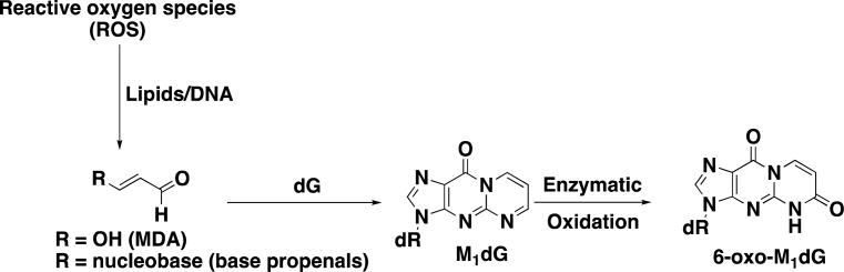
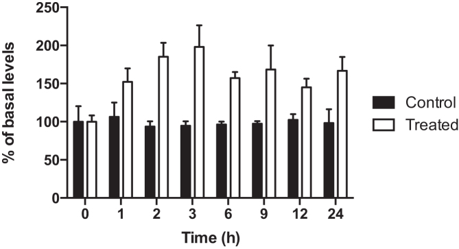
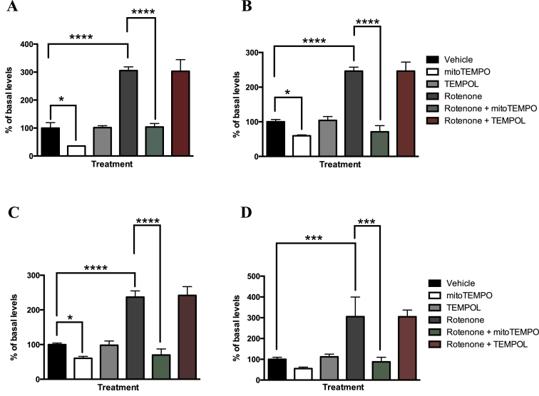
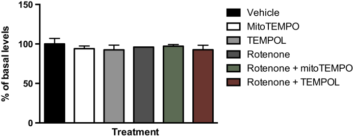
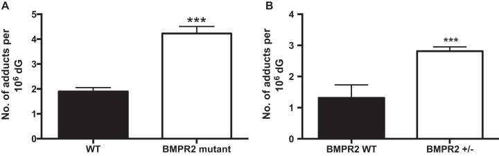
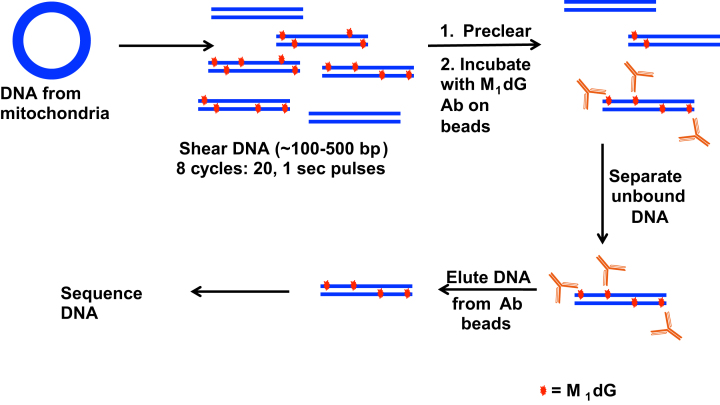
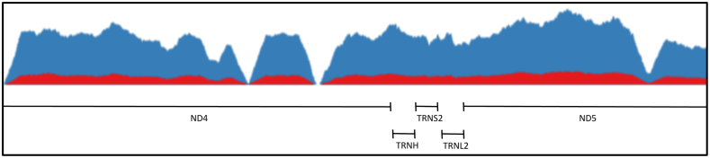
Reactive oxygen species (ROS) are formed in mitochondria during electron transport and energy generation. Elevated levels of ROS lead to increased amounts of mitochondrial DNA (mtDNA) damage. We report that levels of M1dG, a major endogenous peroxidation-derived DNA adduct, are 50-100-fold higher in mtDNA than in nuclear DNA in several different human cell lines. Treatment of cells with agents that either increase or decrease mitochondrial superoxide levels leads to increased or decreased levels of M1dG in mtDNA, respectively. Sequence analysis of adducted mtDNA suggests that M1dG residues are randomly distributed throughout the mitochondrial genome. Basal levels of M1dG in mtDNA from pulmonary microvascular endothelial cells (PMVECs) from transgenic bone morphogenetic protein receptor 2 mutant mice (BMPR2R899X) (four adducts per 106 dG) are twice as high as adduct levels in wild-type cells. A similar increase was observed in mtDNA from heterozygous null (BMPR2+/-) compared to wild-type PMVECs. Pulmonary arterial hypertension is observed in the presence of BMPR2 signaling disruptions, which are also associated with mitochondrial dysfunction and oxidant injury to endothelial tissue. Persistence of M1dG adducts in mtDNA could have implications for mutagenesis and mitochondrial gene expression, thereby contributing to the role of mitochondrial dysfunction in diseases.


Figures

Figure 1.
Figure 1.
Formation of M1dG from MDA and base propenals and the oxidation of M1dG to 6-oxo-M1dG.
Figure 2.
Figure 2.
M1dG levels in mtDNA in RKO cells after treatment with adenine propenal (400 μM) for 1 h. The electrophile was removed, and cells were lysed at the indicated times for mtDNA isolation and analysis. Data represent the mean ± S.D. of triplicate determinations.
Figure 3.
Figure 3.
M1dG levels in the mtDNA of RKO cells (panel A), HEK293 cells (panel B), HepG2 cells (panel C) and RAW264.7 cells (panel D) after treatment with rotenone (100 nM), and/or mitoTEMPO (10 μM), and/or TEMPOL (10 μM) for 24 h. Basal levels of M1dG in mtDNA were determined to be two adducts per 106 dG, 2.3 adducts per 106 dG, two adducts per 106 dG and one adduct per 105 dG in RKO cells, HEK293 cells, HepG2 cells and RAW 264.7 macrophages respectively. Data represent the mean ± S.D. of triplicate determinations.
Figure 4.
Figure 4.
M1dG levels in nuclear DNA in RKO cells after a 24-h treatment with rotenone (100 nM), mitoTEMPO (10 μM), or TEMPOL (10 μM) alone or with rotenone (100 nM) and either mitoTEMPO (10 μM) or TEMPOL (10 μM). Data represent the mean ± S.D. of triplicate determinations. In RKO cells, basal levels of M1dG in genomic DNA was determined to be 1.5 adducts per 108 dG.
Figure 5.
Figure 5.
(A) Mitochondrial M1dG levels in mtDNA in PMVECs isolated from transgenic BMPR2 (bone morphogenetic protein receptor 2) mutant mice (BMPR2 R899X) and wild type mice. Data represent the mean ± S.D. of quadruplicate determinations. (B) Mitochondrial M1dG levels in mtDNA in PMVECs isolated from BMPR2 heterozygous null (BMPR2+/−) cells compared to wild-type. Data represent the mean ± S.D. of quadruplicate determinations.
Figure 6.
Figure 6.
Workflow for the preparation of mtDNA from RKO cells for sequencing analysis following treatment with adenine propenal (400 μM) for 1 h.
Figure 7.
Figure 7.
Representative coverage of a 2 kb region of the mitochondrial genome. The signal corresponding to mtDNA from cells treated with adenine propenal and enriched with the M1dG antibody (blue) shows increased sequence reads but does not show enrichment of distinct regions when compared to the signal corresponding to mtDNA from cells not treated with adenine propenal and enriched (red).

References

    1. Coussens L.M., Werb Z.. Inflammation and cancer. Nature. 2002; 420:860–867. - PMC - PubMed
    1. Elinav E., Nowarski R., Thaiss C.A., Hu B., Jin C., Flavell R.A.. Inflammation-induced cancer: crosstalk between tumours, immune cells and microorganisms. Nat. Rev. Cancer. 2013; 13:759–771. - PubMed
    1. Karin M. NF-kappaB as a critical link between inflammation and cancer. Cold Spring Harb. Perspect. Biol. 2009; 1:a000141. - PMC - PubMed
    1. St-Pierre J., Buckingham J.A., Roebuck S.J., Brand M.D.. Topology of superoxide production from different sites in the mitochondrial electron transport chain. J. Biol. Chem. 2002; 277:44784–44790. - PubMed
    1. Marnett L.J. Oxyradicals and DNA damage. Carcinogenesis. 2000; 21:361–370. - PubMed

Publication types

MeSH terms

Substances

LinkOut - more resources