. 2018 May:518:14-24.
doi: 10.1016/j.virol.2018.02.005. Epub 2018 Feb 12.

Evidence for contemporary plant mitoviruses

Affiliations

Evidence for contemporary plant mitoviruses

Max L Nibert et al. Virology. 2018 May.

Abstract

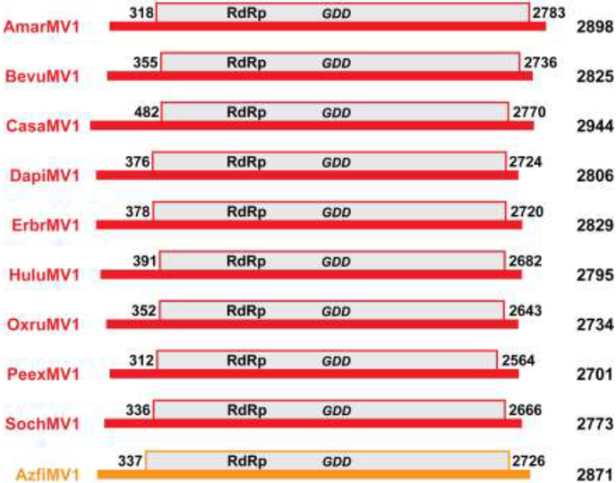
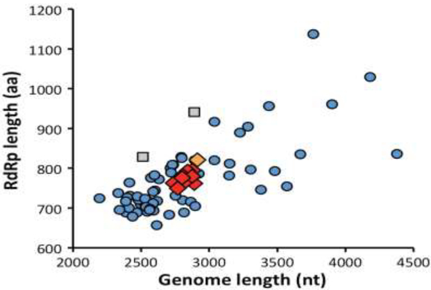
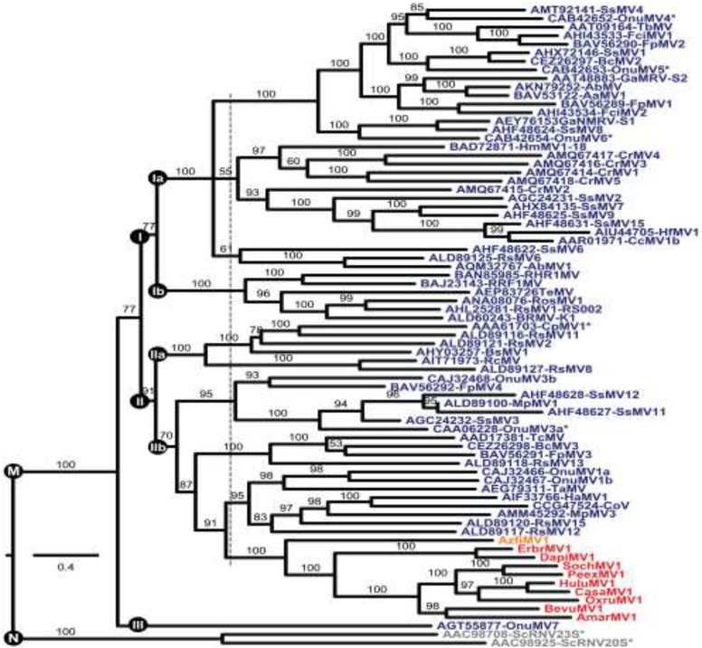
Mitoviruses have small RNA(+) genomes, replicate in mitochondria, and have been shown to infect only fungi to date. For this report, sequences that appear to represent nearly complete plant mitovirus genomes were recovered from publicly available transcriptome data. Twenty of the refined sequences, 2684-2898 nt long and derived from 10 different species of land plants, appear to encompass the complete coding regions of contemporary plant mitoviruses, which furthermore constitute a monophyletic cluster within genus Mitovirus. Complete coding sequences of several of these viruses were recovered from multiple transcriptome (but not genome) studies of the same plant species and also from multiple plant tissues. Crop plants among implicated hosts include beet and hemp. Other new results suggest that such genuine plant mitoviruses were immediate ancestors to endogenized mitovirus elements now widespread in land plant genomes. Whether these mitoviruses are wholly cryptic with regard to plant health remains to be investigated.

Keywords: Database mining; Fungal virus; Mitovirus; Narnaviridae; Plant virus; RNA virus.


Figures

Fig. 1.
Fig. 1.
Scaled diagrams of apparent plant mitovirus genomes. Genome lengths are indicated at right. The genomic RNA plus strand of each virus is shown as a thick horizontal black line. The single long ORF, encoding the viral RdRp, is shown as a gray box above the line. The first and last nt positions in the ORF (including the stop codon) are labeled. The diagrams for the different viruses are aligned according to the position of the conserved GDD motif in each RdRp, which is also labeled. The diagrams for CasaMV1, BevuMV1, DapiMV1, ErbrMV1, and PeexMV1 are those for their reference strains (see Table 1). Color-coding: red, plant mitoviruses from flowering plants (Ambrosia artemisiifolia, Beta vulgaris, Cannabis sativa, Dahlia pinnata, Erigeron breviscapus, Humulus lupulus, Oxybasis rubra, Petunia exserta, and Solanum chacoense); orange, plant mitovirus from a fern (Azolla filiculoides).
Fig. 2.
Fig. 2.
Scatter plot of genome and RdRp lengths. Color-coding for the viruses new to this report (shown as diamonds) is the same as introduced in Fig. 1. Data from previously reported fungal mitoviruses and narnaviruses are shown as blue circles and gray squares, respectively.
Fig. 3.
Fig. 3.
Phylogenetic tree of genus Mitovirus. Deduced RdRp sequences were aligned using MAFFT 7.310 (L-INS-i). The alignment was then analyzed using ModelFinder to determine the best-fit substitution model (VT+F+R6) and subjected to phylogenetic analysis using IQ-TREE and UFBoot as described in Materials and Methods. The site proportions and rates for the FreeRate model in this case were (0.0317,0.0275), (0.0531,0.1920), (0.1140,0.4564), (0.3158,0.8236), (0.3386,1.3151), and (0.1467,1.5775). The tree is displayed as a rectangular phylogram rooted on the branch to genus Narnavirus. Branch support values are shown in %, and branches with <50% support have been collapsed to the preceding node. Scale bar indicates average number of substitutions per alignment position. Color-coding is the same as in Fig. 2. CasaMV1, BevuMV1, DapiMV1, ErbrMV1, and PeexMV1 are represented by their reference strains. Viruses representing the 7 ratified species in genera Mitovirus (M) and Narnavirus (N) are highlighted by asterisks. Three or five apparent main clades within genus Mitovirus are labeled IIII. The dotted vertical line is explained in the main text. See Table S4 for a summary of abbreviations and GenBank numbers.
Fig. 4.
Fig. 4.
Validation results for BevuMV1-VDH66156. Positions of DNA markers are labeled at left (bp). RNA or DNA extracts were obtained from leaves of sugar beet strain VDH66156. (A) RNA extract was used for RT–PCR amplification using three different primer pairs specific for BevuMV1 (V1–V3). (B) RNA extract was used for PCR amplification (no RT step) using two different primer pairs specific for BevuMV1 (V1 and V3). At right is an RT-PCR control using one primer pair specific for BevuMV1 (V1). (C) DNA extract was used for PCR amplification (no RT step) using one primer pair specific for BevuMV1 (V1). At right are PCR controls using two primer pairs specific for B. vulgaris chloroplast DNA (C1 and C2) and two primer pairs specific for B. vulgaris mitochondrial DNA (M1 and M2).
Fig. 5.
Fig. 5.
Phylogenetic tree including plant mitovirus NERVEs. Deduced protein sequences were aligned using MAFFT 7.310 (L-INS-i). The alignment was then analyzed using ModelFinder to determine the best-fit substitution model (VT+F+R6) and subjected to phylogenetic analysis using IQ-TREE and UFBoot as described in Materials and Methods. The site proportions and rates for the FreeRate model in this case were (0.0423,0.0776), (0.0552,0.2609), (0.1204,0.5237), (0.3069,0.8510), (0.3485,1.2689), and (0.1267,1.7035). The tree was displayed as a rectangular phylogram rooted on the branch to genus Narnavirus; the Clade IIb portion (see Fig. 3) was then excerpted for presentation here. Branch support values are shown in %, and branches with <50% support have been collapsed to the preceding node. Scale bar indicates average number of substitutions per alignment position. Color-coding for viruses is the same as in Fig. 2. CasaMV1, BevuMV1, DapiMV1, ErbrMV1, and PeexMV1 are represented by their reference strains. Mitovirus NERVEs are labeled in green; see Table S5 for a summary of their abbreviations and GenBank numbers.

References

    1. Abdoulaye AH, Cheng J, Fu Y, Jiang D, Xie J. 2017. Complete genome sequence of a novel mitovirus from the phytopathogenic fungus Rhizoctonia oryzae-sativae. Arch Virol 162:1409–1412. - PubMed
    1. Ballinger MJ, Bruenn JA, Taylor DJ. 2012. Phylogeny, integration and expression of sigma virus-like genes in Drosophila. Mol Phylogenet Evol 65:251–258. - PubMed
    1. Bartholomäus A, Wibberg D, Winkler A, Pühler A, Schlüter A, Varrelmann M. 2016. Deep sequencing analysis reveals the mycoviral diversity of the virome of an avirulent isolate of Rhizoctonia solani AG-2–2 IV. PLoS One 11:e0165965. - PMC - PubMed
    1. Bruenn JA, Warner BE, Yerramsetty P. 2015. Widespread mitovirus sequences in plant genomes. PeerJ. 3:e876. - PMC - PubMed
    1. Brundrett MC. 2002. Coevolution of roots and mycorrhizas of land plants. New Phytol 154: 275–304. - PubMed

Publication types

MeSH terms