. 2018 Mar 16;359(6381):1274-1277.
doi: 10.1126/science.aao6891. Epub 2018 Feb 8.

Rev-erbα dynamically modulates chromatin looping to control circadian gene transcription

Affiliations

Rev-erbα dynamically modulates chromatin looping to control circadian gene transcription

Yong Hoon Kim et al. Science. .

Abstract

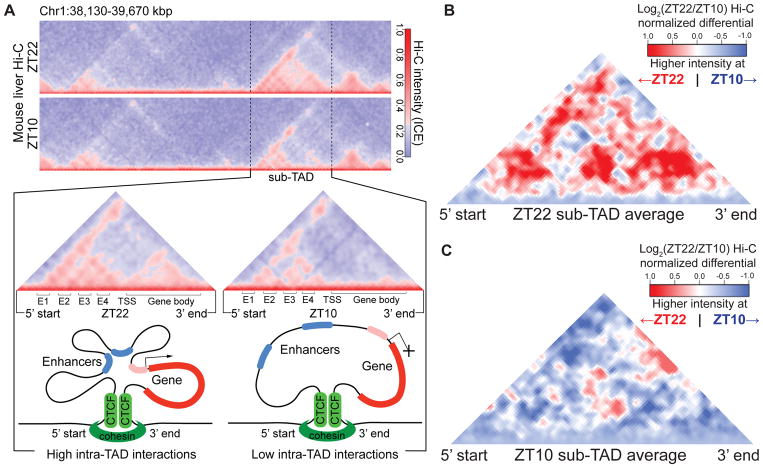
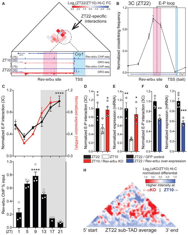
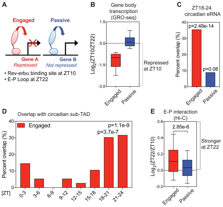
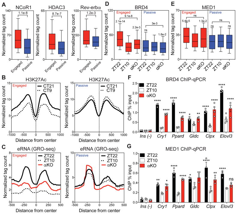
Mammalian physiology exhibits 24-hour cyclicity due to circadian rhythms of gene expression controlled by transcription factors that constitute molecular clocks. Core clock transcription factors bind to the genome at enhancer sequences to regulate circadian gene expression, but not all binding sites are equally functional. We found that in mice, circadian gene expression in the liver is controlled by rhythmic chromatin interactions between enhancers and promoters. Rev-erbα, a core repressive transcription factor of the clock, opposes functional loop formation between Rev-erbα-regulated enhancers and circadian target gene promoters by recruitment of the NCoR-HDAC3 co-repressor complex, histone deacetylation, and eviction of the elongation factor BRD4 and the looping factor MED1. Thus, a repressive arm of the molecular clock operates by rhythmically modulating chromatin loops to control circadian gene transcription.


Figures

Figure 1
Figure 1. Circadian sub-TADs undergo rhythmic intra-TAD compaction within stable boundaries
(A) Heat maps of ZT22 and ZT10 Hi-C demonstrating circadian intra-TAD interactions within sub-TAD boundaries (dotted lines), as represented by Hi-C intensity normalized by Iterative Correction and Eigenvector decomposition (ICE). Transcriptional start site of the Npas2 gene (TSS) forms rhythmic intra-TAD loops with upstream enhancers (E1-4) as well as with the gene body, as illustrated by schematics below. (B) ZT22 sub-TAD and (C) ZT10 sub-TAD averaged differential changes in intra-TAD interactions visualized as log2 ratio within size-normalized sub-TAD 5’ and 3’ boundaries (red= higher interaction ratio at ZT22, blue=higher interaction ratio at ZT10).
Figure 2
Figure 2. Rev-erbα causally opposes enhancer-promoter loop formation
(A) Differential Hi-C analysis at the Cry 1 locus revealing ZT22-specific interactions, represented as log2 ratio (ZT22 Hi-C/ZT10 Hi-C). ZT22-specific interactions (dotted circle) occur between a region around the intronic Rev-erbα site (red) and the Cry1 TSS (blue). Global Run-On seq (GRO-seq) demonstrates circadian nascent transcription as well as the presence of bidirectional eRNA at Rev-erbα site at ZT22. (B) 3C validation of enhancer-promoter loop (E-P loop) identified at ZT22 between Rev-erbα site (red) and TSS (blue) (n=5, mean ±SEM). (C) Circadian plot demonstrating Cry1 E-P loop, mRNA expression, and Rev-erbα ChIP ±SEM (n=4–5, p values shown for 3C and ChIP peaks compared to troughs, one-way ANOVA followed by multiple comparisons correction with the Tukey method) (D) E-P loop and (E) mRNA expression of Cry1 at ZT22 (black), ZT10 (white), and ZT10 Rev-erbα KO (red), represented as mean ±SEM (n=4, one-way ANOVA followed by Dunnett’s multiple comparisons test). (F) E-P loop and (G) mRNA expression of Cry1 at ZT22 with control GFP expression (black) versus Rev-erbα over-expression (blue) expressed as mean ±SEM (n=5, two-tailed Student’s t-test). (H) Same analysis as in Fig. 1B, but comparing ZT10 Rev-erbα KO (αKO) to ZT10 WT at ZT22 sub-TADs (red= higher interaction ratio at ZT10 αKO, blue=higher interaction ratio at ZT10 WT). * p<0.05, ** p<0.01, *** p<0.001, **** p<0.0001
Figure 3
Figure 3. Rev-erbα attenuates enhancer-promoter looping at functional binding sites
(A) Rev-erbα sites at E-P loops were classified as “engaged” when looped to genes whose transcription was repressed at ZT10 relative to ZT22, and otherwise classified as “passive”. (B) Gene body transcription change between ZT22 and ZT10 at engaged vs. passive sites. Engaged target genes were defined based on nascent gene body transcription fold change ≥1.5 between ZT22 and ZT10. (C) Engaged Rev-erbα sites were highly correlated with ZT18-24 circadian eRNAs (6) (within ± 2kbp, one-tailed hypergeometric tests). (D) Engaged Rev-erbα sites were confined within sub-TADs that contain circadian genes peaking at ZT18-24 (one-tailed hypergeometric tests). (E) E-P loops between engaged Rev-erbα binding sites and target gene promoters were stronger at ZT22 than ZT10 (Mann-Whitney test). For boxplots, whiskers drawn at 10th and 90th percentiles
Figure 4
Figure 4. Functional Rev-erbα binding evicts BRD4 and MED1 from sites of looping
(A) Higher recruitment of NCoR1 and HDAC3 at engaged Rev-erbα sites associated with a slight average increase in Rev-erbα binding (Mann-Whitney tests). (B) Circadian deacetylation of histone 3 lysine 27 (H3K27Ac) at circadian time 21 (CT21, black) and CT9 (dotted) at engaged vs. passive sites. (C) Circadian eRNA transcription between ZT22 (black) and ZT10 (dotted), with increased transcription at ZT10 in αKO (red) at engaged sites. (D) Circadian eviction of BRD4 and (E) MED1 between ZT22 and ZT10, with enhanced binding at ZT10 in αKO at engaged sites (Dunn’s multiple comparisons tests after one-way ANOVA/Friedman test). (F) ChIP-qPCR validation of BRD4 and (G) MED1 eviction at ZT10 and enhanced binding at ZT10 in αKO at engaged sites (Ins as a negative control, n=3–4, two-way ANOVA followed by Dunnett’s multiple comparisons test). For boxplots, whiskers drawn at 10th and 90th percentiles, ns p≥0.05, * p<0.05, ** p<0.01, *** p<0.001, **** p<0.0001

Comment in

  • Circadian organization of the genome.
    Mallet de Lima CD, Göndör A. Mallet de Lima CD, et al. Science. 2018 Mar 16;359(6381):1212-1213. doi: 10.1126/science.aat0934. Science. 2018. PMID: 29590061 No abstract available.

References

    1. Bass J, Lazar MA. Circadian time signatures of fitness and disease. Science. 2016;354:994–998. - PubMed
    1. Takahashi JS. Transcriptional architecture of the mammalian circadian clock. Nat Rev Genet. 2016;18:164–179. - PMC - PubMed
    1. Feng D, et al. A Circadian rhythm orchestrated by histone deacetylase 3 controls Hepatic lipid metabolism. Science. 2011;331:1315–1319. - PMC - PubMed
    1. Koike N, et al. Transcriptional architecture and chromatin landscape of the core circadian clock in mammals. Science. 2012;338:349–54. - PMC - PubMed
    1. Vollmers C, et al. Circadian oscillations of protein-coding and regulatory RNAs in a highly dynamic mammalian liver epigenome. Cell Metab. 2012;16:833–845. - PMC - PubMed

Publication types

MeSH terms