Review
. 2018 Mar;103(3):382-394.
doi: 10.3324/haematol.2017.183335. Epub 2018 Feb 8.

Extracellular vesicles in the hematopoietic microenvironment

Affiliations
Review

Extracellular vesicles in the hematopoietic microenvironment

John T Butler et al. Haematologica. 2018 Mar.

Abstract

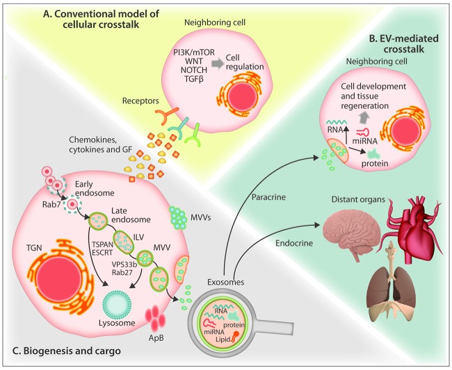
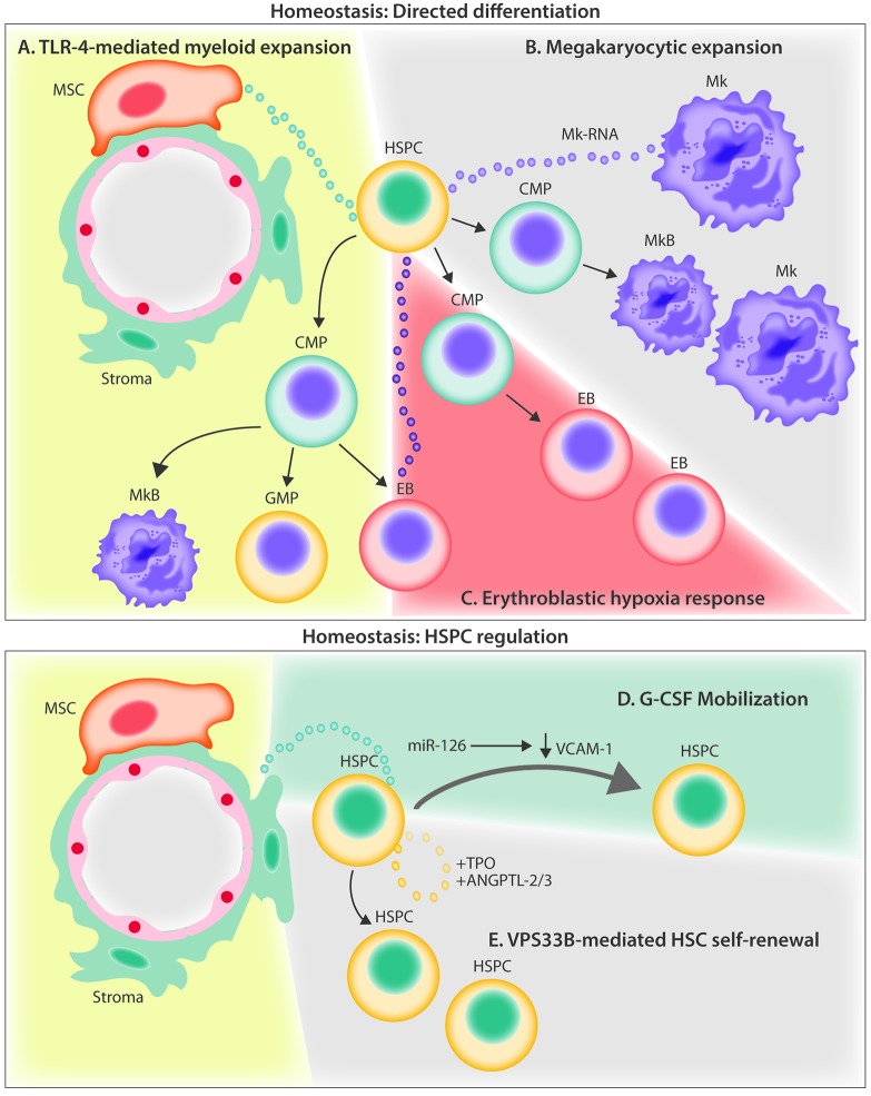
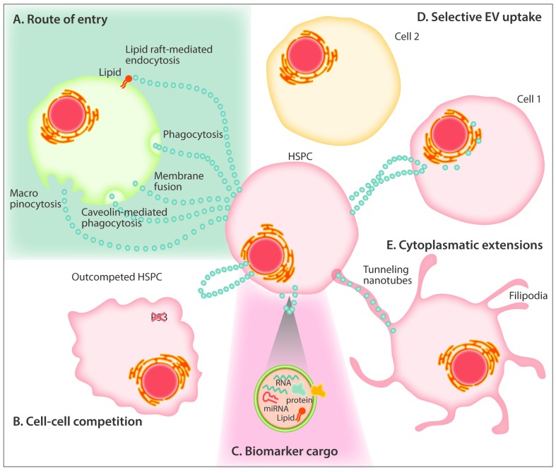
Self-renewal and differentiation are defining characteristics of hematopoietic stem and progenitor cells, and their balanced regulation is central to lifelong function of both blood and immune systems. In addition to cell-intrinsic programs, hematopoietic stem and progenitor cell fate decisions are subject to extrinsic cues from within the bone marrow microenvironment and systemically. Yet, many of the paracrine and endocrine mediators that shape hematopoietic function remain to be discovered. Extracellular vesicles serve as evolutionarily conserved, constitutive regulators of cell and tissue homeostasis, with several recent reports supporting a role for extracellular vesicles in the regulation of hematopoiesis. We review the physiological and pathophysiological effects that extracellular vesicles have on bone marrow compartmental function while highlighting progress in understanding vesicle biogenesis, cargo incorporation, differential uptake, and downstream effects of vesicle internalization. This review also touches on the role of extracellular vesicles in hematopoietic stem and progenitor cell fate regulation and recent advances in therapeutic and diagnostic applications of extracellular vesicles in hematologic disorders.


Figures

Figure 1.
Figure 1.
Schematic representation of biogenesis of extracellular vesicles and unique aspects of their trafficking. (A) The conventional model of cellular crosstalk involves receptor-ligand interactions between secreted chemokines, cytokines and growth factors and cellular surface receptors. (B) EV-mediated crosstalk occurs through the trafficking of vesicle-associated protein, lipid and RNA components to proximal cells or to distal organs via the bloodstream in a “paracrine” or “endocrine” manner, respectively. (C) Exosomes are formed from the maturation of early endosomes into Rab7-containing late endosomes leading to the generation of intraluminal vesicles via the action of tetraspanin and ESCRT proteins which sort the endosomal constituents into distinct multivesicular bodies. Through the action of Rab27 and VPS33b, multivesicular bodies evade lysosome degradation and fuse with the plasma membrane to release 30–125 nm exosomes. Cells also release 50–1000 nm microvesicles that form through calcium-mediated budding of the plasma membrane, and during programed cell death, large (>1000 nm) apoptotic bodies. ApB: apoptotic bodies ESCRT: endosomal-sorting complex required for transport; GF: growth factors; ILV: intraluminal vesicle; MV: microvesicle; MVB: multivesiclular bodies; mTOR: mammalian target of rapamycin; PI3K; phosphatidylinositol-3 kinase; TGF-β: transforming growth factor beta; TGN: trans-Golgi network; TSPAN: tetraspanin; VPS33B: vacuolar protein sorting-associated protein 33B.
Figure 2.
Figure 2.
Current evidence for extracellular vesicle crosstalk in the homeostatic bone marrow microenvironment. (A) MSC-derived EVs signal to HSPCs through the TLR-4 pathway, resulting in myeloid biased expansion. (B) Megakaryocyte-derived MVs are internalized by HSPCs and increase differentiation of new megakaryocytes through RNA-mediated signaling. (C) Hypoxia induces erythroleukemia cells to release EVs containing miR-486 which increases erythroblastic differentiation by targeting Sirt1 in HSPCs. (D) G-CSF infusion stimulates the release of EVs containing miR-126 that act to down-regulate VCAM-1 in HSPCs, resulting in their mobilization out of the BM. (E) HSPCs autoregulate stem potential by packaging and releasing critical secretory proteins through the exosomal pathway via the action of VPS33B. ANGPTL-2/3; angiopoietin-like protein 2 and 3; BM: bone marrow; CMP: common myeloid progenitor; EB: erythroblast; EVs: extracellular vesicles; G-CSF: granulocyte colony-stimulating factor; GMP; granulocyte monocyte progenitor; HSPC: hematopoietic stem and progenitor cell; Mk: megakaryocytes; MkB: megakaryoblast; MSC: mesenchymal stem cell; miR: microRNA; MV: microvesicles; TLR-4: Toll-like receptor 4; TPO: thrombopoietin; VCAM-1: vascular cell adhesion molecule; VPS33B: vacuolar protein sorting-associated protein 33B.
Figure 3.
Figure 3.
Current evidence for extracellular vesicle crosstalk in the leukemic microenvironment (A) EVs from AML blasts traffic miR-155 to HSPCs and down-regulate critical transcription factor, c-MYB, resulting in reduced differentiation potential. (B) AML EVs reprogram MSCs and stromal cells, and downregulate niche retention factor CXCL12 resulting in mobilization of HSPCs from the BM. (C) AML and MDS EVs promote the loss of HSPC supportive factors, CXCL12, SCF, IGF-1 through the trafficking of miR-7797 to supportive stroma, leading to reduced HSPC viability and hematopoietic potential. AML: acute myelogenous leukemia; ANGPT-1: angiopoietin 1; BM: bone marrow; CXCL12: C-X-C motif chemokine 12; EVs: extracellular vesicles; HSPCs: hematopoietic stem and progenitor cells; IGF-1: insulin-like growth factor 1; MDS: myelodysplastic syndrome; miR: microRNA; MSC: mesenchymal stem cell; PCBP1: poly(rc) binding protein 1; SCF: stem cell factor.
Figure 4.
Figure 4.
Unresolved aspects of extracellular vesicle biology in the regulation of hematopoiesis. (A) EVs have been proposed to enter recipient cells through lipid raft-mediated internalization, endocytosis, phagocytosis, membrane fusion, caveolin-mediated endocytosis and macropinocytosis. (B) Exosome-mediated crosstalk may explain the intercellular competition of neighboring cells where the “winner” HSPC outcompetes the less fit HSPC through a P53-dependent mechanism. (C) Vesicles contain cargo comprised of uniquely packaged proteins, miRNAs and RNAs which serve as promising biomarkers for disease detection. (D) Vesicles from HSPCs and other cells of the bone marrow niche have been shown to exhibit preferential targeting to specific recipient cells for entry. (E) Cytonemes (filopodia, invadopodia, tunneling nanotubes) are cytoplasmic extensions that serve as modes of exosomal transfer to adjacent bystander cells. EVs: extracellular vesicles; HSPC: hematopoietic stem and progenitor cell.

References

    1. Morrison SJ, Scadden DT. The bone marrow niche for haematopoietic stem cells. Nature. 2014;505(7483):327–334. - PMC - PubMed
    1. Pietras EM, Warr MR, Passegué E. Cell cycle regulation in hematopoietic stem cells. J Cell Biol. 2011;195(5):709–720. - PMC - PubMed
    1. Schepers K, Campbell TB, Passegué E. Normal and leukemic stem cell niches: insights and therapeutic opportunities. Cell Stem Cell. 2015;16(3):254–267. - PMC - PubMed
    1. Blank U, Karlsson S. TGF-β signaling in the control of hematopoietic stem cells. Blood. 2015;125(23):3542–3550. - PubMed
    1. Yamazaki S, Iwama A, Takayanagi S-I, et al. Cytokine signals modulated via lipid rafts mimic niche signals and induce hibernation in hematopoietic stem cells. EMBO. 2006;25(15):3515–3523. - PMC - PubMed

Publication types