Review
. 2018 Jun;70(6):805-816.
doi: 10.1002/art.40452. Epub 2018 Apr 18.

Review: Cell Death, Nucleic Acids, and Immunity: Inflammation Beyond the Grave

Affiliations
Review

Review: Cell Death, Nucleic Acids, and Immunity: Inflammation Beyond the Grave

Keith B Elkon. Arthritis Rheumatol. 2018 Jun.

Abstract

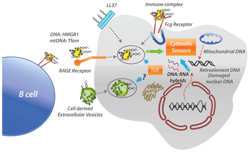
Cells of the innate immune system are rigged with sensors that detect nucleic acids derived from microbes, especially viruses. It has become clear that these same sensors that respond to nucleic acids derived from damaged cells or defective intracellular processing are implicated in triggering diseases such as lupus and arthritis. The ways in which cells die and the concomitant presence of proteins and peptides that allow nucleic acids to re-enter cells profoundly influence innate immune responses. In this review, we briefly discusses different types of programmed necrosis, such as pyroptosis, necroptosis, and NETosis, and explains how nucleic acids can engage intracellular receptors and stimulate inflammation. Host protective mechanisms that include compartmentalization of receptors and nucleases as well as the consequences of nuclease deficiencies are explored. In addition, proximal and distal targets in the nucleic acid stimulation of inflammation are discussed in terms of their potential amenability to therapy for the attenuation of innate immune activation and disease pathogenesis.


Figures

Figure 1
Figure 1. Basic concepts of Innate cell stimulation by
Damage (DAMPs) or Pathogen (PAMPs) Associated Molecular Patterns. Either DAMPs or PAMPs are capable of activating cells of the innate immune system such as macrophages and dendritic cells. Depending on the nature of molecular pattern, either anti- or pro-inflammatory responses can be elicited. These responses impact both the inflammatory environment as well as communication with T cells as shown in the figure. Whereas intact apoptotic cells are immunologically silent or stimulate anti-inflammatory responses as well as negative adaptive responses (as in the left panel), release of cellular content from necrotic cells is almost always inflammatory (as in the right panel). Note that DAMPs and PAMPs also impact B cells both by serving as antigens activating the B cell receptor and, following internalization, by activation of sensors such as TLR 7 and 9 (see also Fig. 3).
Figure 2
Figure 2. Immediate or Programmed forms of necrosis stimulate inflammation
Programmed forms of necrosis include pyroptosis, necroptosis and NET formation – see text for details. Common features are damage to the cell membrane and release of cytokines, although this is less well established for NET formation. Gasdermin-D (GSDMD) and mixed lineage kinase-domain like (MLKL) are proteins involved in pore formation in pyroptosis and necroptosis respectively. How chromatin escapes from neutrophils during NET formation is unknown.
Figure 3
Figure 3. Nucleic acids gain access to intracellular sensors through multiple routes
Extracellular nucleic acids (DNA or RNA) can be captured by autoantibodies, bind to highly cationic peptides such as LL-37 or interact with nucleic acid binding proteins such as HMGB1 to gain re-entry into cells. A professional phagocytic cell such as the macrophage depicted on the right may uptake molecules by all of these pathways, whereas in other cell types, uptake may be more restricted. Endocytosis of nucleic acid complexes may be receptor mediated (Fcg receptor and RAGE receptors are shown) but in other cases, mechanisms are less clear as discussed in the text. Nucleic acid complexes that enter endosomes generally trigger TLRs but escape into the cytosol has been documented in many situations (green arrow). Intracellular nucleic acids gain access to sensors through different mechanisms – mitochondrial DNA may be released directly from the damaged organelle or renter the cell after binding to the protein called Tfam. The cytosolic sensors can be activated by DNA:RNA hybrids as well as nuclear DNA arising from retroelements or nuclear damage. Note that cell debris, especially highly charged DNA and RNA may also engage and be internalized by the B cell receptor thereby delivering to the B cell signals through BCR as well as TLR.
Figure 4
Figure 4. Sensors of intracellular nucleic acids
Intracellular receptors responsive to DNA (black) and RNA (red) are shown. The subset of nucleic acid responsive TLRs are present in the endosomal compartment whereas most other sensors are present in the cytosol. The signal transduction and also the transcription pathways are simplified. Adapters / platforms are shown in blue, red arrows indicate phosphorylation reactions; transcription factors are shaded in yellow. Abbreviations: AIM2, absent in melanoma-2; ALRs, AIM2-like receptors;; cGAMP, cyclic GMP–AMP; cGAS, cGAMP synthase; ER, endoplasmic reticulum; IFN, interferon; IKK (IKB kinase); IL, interleukin; IRAK (interleukin-1 receptor-associated kinase 4), IRF, IFN regulatory factor; MyD88, myeloid differentiation primary response protein-88; NALP3, NACHT, LRR and PYD domains-containing protein 3; PRR, pattern recognition receptor; RIG-I, retinoic acid–inducible gene I; STING, stimulator of IFN genes; TBK (TANK binding kinase); TLR, Toll-like receptor.
Figure 5
Figure 5. Potential points of therapeutic intervention in response to nucleic acid stimulation of sensors
Biologic or small molecule drugs have been used in patients or in preclinical models to block cytokines (#2) or downstream signaling events (#1). Antibodies targeting plasmacytoid dendritic cells are in early clinical trials (#3). The intracellular TLR sensors can be inhibited directly by oligonucleotides (ODN) or TLR signaling can be modulated by chemical inhibitors of IRF5 or IRAK4 (#4). Similarly, cGAS-STING activation or signaling can be attenuated by antimalarial drugs or TBK1 inhibitors (#5). Nucleases like DNase and RNase (#6) can degrade nucleic acid antigens that are free, in immune complexes or released by NET formation. Inhibitors of PAD4 or mitochondrial ROS inhibit the process of NET formation (#7). See text for details.

References

    1. Watanabe-Fukunaga R, Brannan CI, Copeland NG, Jenkins NA, Nagata S. Lymphoproliferation disorder in mice explained by defects in Fas antigen that mediates apoptosis. Nature. 1992;356:314–7. - PubMed
    1. Casciola-Rosen LA, Anhalt G, Rosen A. Autoantigens targeted in systemic lupus erythematosus are clustered in two populations of surface structures on apoptotic keratinocytes. Journal of Experimental Medicine. 1994;179:1317–30. - PMC - PubMed
    1. Martin SJ, Henry CM, Cullen SP. A perspective on mammalian caspases as positive and negative regulators of inflammation. Mol Cell. 2012;46(4):387–97. - PubMed
    1. Janeway CA., Jr Approaching the asymptote? Evolution and revolution in immunology. Cold Spring Harb Symp Quant Biol. 1989;54(Pt 1):1–13. - PubMed
    1. Matzinger P. The danger model: a renewed sense of self. Science. 2002;296(5566):301–5. - PubMed

Publication types