. 2018 Jun;70(6):855-867.
doi: 10.1002/art.40447. Epub 2018 Apr 18.

Augmented Th17 Differentiation Leads to Cutaneous and Synovio-Entheseal Inflammation in a Novel Model of Psoriatic Arthritis

Affiliations

Augmented Th17 Differentiation Leads to Cutaneous and Synovio-Entheseal Inflammation in a Novel Model of Psoriatic Arthritis

Lu Yang et al. Arthritis Rheumatol. 2018 Jun.

Abstract

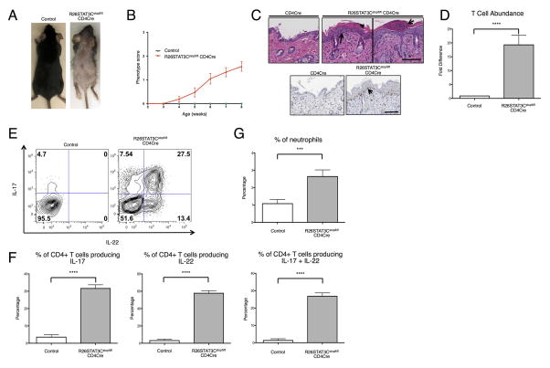
Objective: To introduce a novel preclinical animal model of psoriatic arthritis (PsA) in R26Stat3Cstopfl/fl CD4Cre mice, and to investigate the role of Th17 cytokines in the disease pathogenesis.

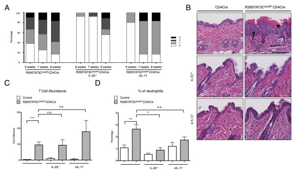
Methods: We characterized a novel murine model of Th17-driven cutaneous and synovio-entheseal disease directed by T cell-specific expression of a hyperactive Stat3 allele. By crossing R26Stat3Cstopfl/fl CD4Cre mice onto an interleukin-22 (IL-22)-knockout background or treating the mice with a neutralizing antibody against IL-17, we interrogated how these Th17 cytokines could contribute to the pathogenesis of PsA.

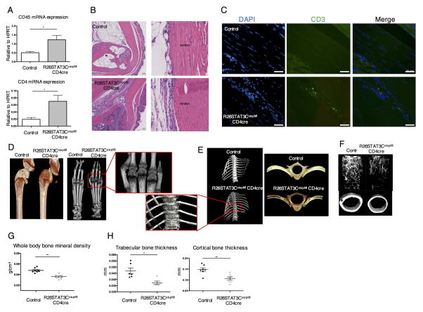
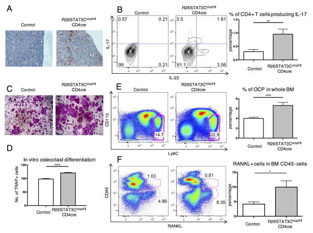
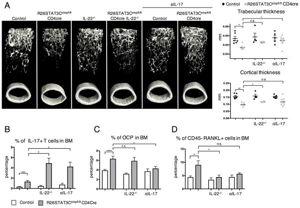
Results: R26Stat3Cstopfl/fl CD4Cre mice developed acanthosis, hyperkeratosis, and parakeratosis of the skin, as well as enthesitis/tendinitis and periarticular bone erosion in different joints, accompanied by osteopenia. T cell-specific expression of a hyperactive Stat3C allele was found to drive the augmented Th17 response in these animals. Careful characterization of the mouse bone marrow revealed an increase in osteoclast progenitor (OCP) and RANKL-producing cells, which contributed to the osteopenia phenotype observed in the mutant animals. Abrogation of the Th17 cytokines IL-17 or IL-22 improved both the skin and bone phenotype in R26Stat3Cstopfl/fl CD4Cre mice, revealing a central role of Th17 cells in the regulation of OCP and RANKL expression on stromal cells.

Conclusion: Perturbation of the IL-23/Th17 axis instigates Th17-mediated inflammation in R26Stat3Cstopfl/fl CD4Cre mice, leading to cutaneous and synovio-entheseal inflammation and bone pathologic features highly reminiscent of human PsA. Both IL-17A and IL-22 produced by Th17 cells appear to play critical roles in promoting the cutaneous and musculoskeletal inflammation that characterizes PsA.


Figures

Figure 1
Figure 1. R26STAT3Cstopfl/fl CD4Cre mice developed a psoriatic-like skin phenotype
(A) Representative image of 8-week-old R26STAT3Cstopfl/fl CD4Cre (skin score 3) and littermate R26STAT3Cstopfl/fl control mice. (B) Average skin phenotype scores of R26STAT3Cstopfl/fl CD4Cre (n=61) and control (n=20) mice from 2 to 8 weeks of age. The scale ranges from 0 (no phenotype) to 3 (greater than 50% fur loss and/or visibly dry/crusty skin). See methods for detailed description of the scoring system. (C) Representative histological skin sections from 8–week-old control and R26STAT3Cstopfl/flCD4Cre mice. Scale bar = 100 μm. Top middle panel: H&E staining. Arrow points to acanthosis and arrowhead points to parakeratosis in an area of hyperkeratosis. Top right panel: H&E staining. Arrow indicates an area reminiscent of Munro’s abscess. Bottom: Anti-CD3 staining, arrow highlights T cells tracking the epidermal/dermal boundary. (D) Fold difference of CD3+CD4+ T cells isolated from skin of R26STAT3Cstopfl/fl CD4Cre and control mice. (E) Representative intracellular flow cytometry analysis from CD3+CD4+ T cells isolated from the skin of 6–10 weeks old R26STAT3Cstopfl/fl CD4Cre and control mice. (F) Percentage of CD3+CD4+ skin T cells expressing IL-17 (left), IL-22 (middle), or both (right). (G) Percentage of neutrophils in the skin. For D–G, data is representative of 15 independent experiments with n ≥17 for each genotype, aged 6–10 weeks old. Statistical significance was assessed using (D) the Sign test and (F–G) the Mann-Whitney U test. Significance values are as follows: *** p ≤ 0.001; **** p ≤ 0.0001.
Figure 2
Figure 2. R26STAT3Cstopfl/fl CD4Cre mice presented tendonitis/enthesitis, periarticular bone erosion and osteopenia phenotype
(A) Quantitative PCR analysis of CD45 and CD4 expression in mRNA isolated from Achilles tendon and entheseal tissues of R26STAT3Cstopfl/fl CD4Cre mice and their littermate controls, (R26STAT3Cstopfl/+ or R26STAT3Cstopfl/fl mice). (B) Representative H&E staining sections of the ankle joints from control and R26STAT3Cstopfl/fl CD4Cre mice. (B) scale bar = 100μm. (C) Representative examples of immunofluorescent staining images of Achilles tendon from control and R26STAT3Cstopfl/fl CD4Cre mice. (C) scale bar = 50μm. Representative microCT images of knees (D), paws (D), vertebra (E), and femurs (F) of R26STAT3Cstopfl/fl CD4Cre mice and their littermate controls. (G) Whole body bone mineral density of control and R26STAT3Cstopfl/fl CD4Cre mice measured by DEXA scan. (H) MicroCT analysis on femur trabecular and cortical thickness of control and R26STAT3Cstopfl/fl CD4Cre mice. Each point in figure G and H represents one animal. Analysis was performed on mice aged between 6 to 10 weeks old. Number of independent experiments ≥ 3. Statistical significance was assessed using the nonparametric two-tailed Mann-Whitney U test. Significance values are as follows: ns p > 0.05; *p ≤ 0.05; ** p ≤ 0.01.
Figure 3
Figure 3. Th17-driven osteopenia phenotype of R26STAT3Cstopfl/fl CD4Cre mice was correlated with an increase in osteoclast progenitor cells and RANKL+ cells in the local environment
(A) Representative immunohistochemistry staining of CD3 on the femur of control and R26STAT3Cstopfl/fl CD4Cre mice. (B) Representative intracellular staining of IL-17 and IL-22 in CD3+ CD4+ T cells in the bones of control and R26STAT3Cstopfl/fl CD4Cre mice (left) and quantified percentage of IL-17 producers in CD3+ CD4+ T cells (right). (C) Representative images of TRAP stained osteoclasts following in vitro osteoclast differentiation of bone marrow cells from R26STAT3Cstopfl/fl CD4Cre mice and their littermate controls. (D) Number of TRAP+ cells at day 10 of an in vitro osteoclast differentiation protocol normalized to number of TRAP+ cells derived from control bone marrow. (E–F) Flow cytometric analysis of OCP population and RANKL producers in the bone marrow of R26STAT3Cstopfl/fl CD4Cre mice and their littermate controls. All analysis was performed on mice aged between 6 to 10 weeks. Number of independent experiments ≥ 3 for all assays. Statistical significance was assessed using the nonparametric two-tailed Mann-Whitney U test. Significance values are as follows: ns p > 0.05; ** p ≤ 0.01; *** p ≤ 0.001; **** p ≤ 0.0001.
Figure 4
Figure 4. Genetic ablation of IL-22 and αIL-17 Ab treatment led to amelioration of the psoriatic skin phenotype in R26STAT3Cstopfl/fl CD4Cre mice
(A) Phenotype score of R26STAT3Cstopfl/fl CD4Cre mice, R26STAT3Cstopfl/fl CD4Cre IL-22−/− mice, and R26STAT3Cstopfl/fl CD4Cre αIL-17 treated mice at 6, 7, and 8 weeks of age. Data represents 10 to 21 mice per group. The scale ranges from 0 (no phenotype) to 3 (greater than 50% fur loss and/or visibly dry/crusty skin). See methods for detailed description of the scoring system. (B) Representative H&E staining of skin sections from control and R26STAT3Cstopfl/fl CD4Cre mice. Mice are 8 weeks old. Scale bar = 100 μm. Top panel: skin from untreated mice. Middle panel: skin from mice on an IL-22 knockout background. Bottom panel: skin from αIL-17 treated mice. (C) Fold difference of CD3+CD4+ T cells from the skin of R26STAT3Cstopfl/fl CD4Cre and control mice, normalized to the untreated controls within each experiment. (D) Percentage of neutrophils from skin of R26STAT3Cstopfl/fl CD4Cre and control mice. Mice are untreated controls, IL-22−/−, or αIL-17 treated. For C–D Data is representative of n ≥ 3 independent experiments with n ≥ 6 for each genotype, 6–10 weeks old. Statistical significance was assessed using the (C) Sign test and (C–D) the nonparametric two-tailed Mann-Whitney U test. Significance values are as follows: ns p > 0.05; ** p ≤ 0.01; *** p ≤ 0.001; **** p ≤ 0.0001.
Figure 5
Figure 5. IL-22 deficiency and αIL-17 treatment partially ameliorated the bone defects in R26STAT3Cstopfl/fl CD4Cre mice
(A) MicroCT analysis on the femur of R26STAT3Cstopfl/fl CD4Cre mice and their littermate controls that were crossed onto IL-22−/− background or treated with αIL-17. The 3D images showing femur bones of these mice (left) with trabecular and cortical thickness of these animals quantified (right). (B) Percentage of CD3+ CD4+ cells that produce IL-17 in the bone marrow of R26STAT3Cstopfl/fl CD4Cre mice and their littermate controls that were crossed onto IL-22−/− background or treated with αIL-17. (C–D) Percentage of osteoclast progenitor cells and RANKL producers in the bone marrow of IL-22−/− and αIL-17 treated mice. Analysis was performed on mice aged between 6 to 10 weeks old. Number of independent experiments ≥ 3 for all assays. Statistical significance was assessed using the nonparametric two-tailed Mann-Whitney U test. Significance values are as follows: ns p > 0.05; ** p ≤ 0.01; *** p ≤ 0.001; **** p ≤ 0.0001.

Comment in

References

    1. Moll J, Wright V. Seminars in arthritis and rheumatism. Elsevier; 1973. Psoriatic arthritis. - PubMed
    1. Ritchlin CT, Colbert RA, Gladman DD. Psoriatic Arthritis. New England Journal of Medicine. 2017;376(10):957–970. - PubMed
    1. Cantini F, et al. Psoriatic arthritis: a systematic review. Int J Rheum Dis. 2010;13(4):300–17. - PubMed
    1. Gladman D, et al. Psoriatic arthritis: epidemiology, clinical features, course, and outcome. Annals of the rheumatic diseases. 2005;64(suppl 2):ii14–ii17. - PMC - PubMed
    1. Ogdie A, et al. The risk of fracture among patients with psoriatic arthritis and psoriasis: a population-based study. Annals of the Rheumatic Diseases. 2017 p. annrheumdis-2016-210441. - PMC - PubMed

Publication types