. 2018 Feb 12;5(1):13.
doi: 10.3390/jcdd5010013.

Hyperglycemia Alters the Structure and Hemodynamics of the Developing Embryonic Heart

Affiliations

Hyperglycemia Alters the Structure and Hemodynamics of the Developing Embryonic Heart

Taylor B Lawson et al. J Cardiovasc Dev Dis. .

Abstract

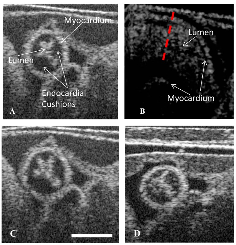
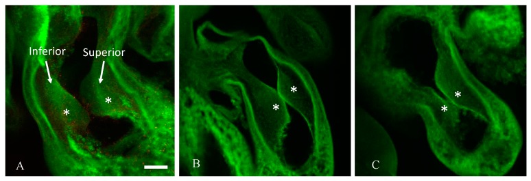
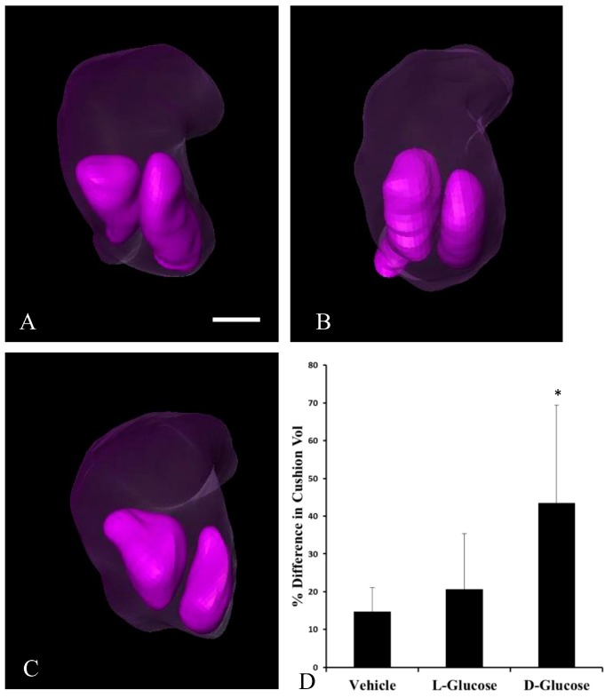
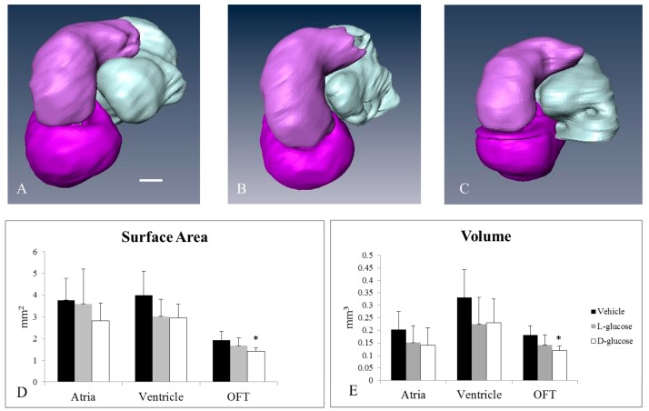
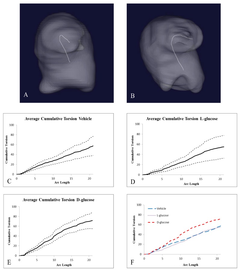
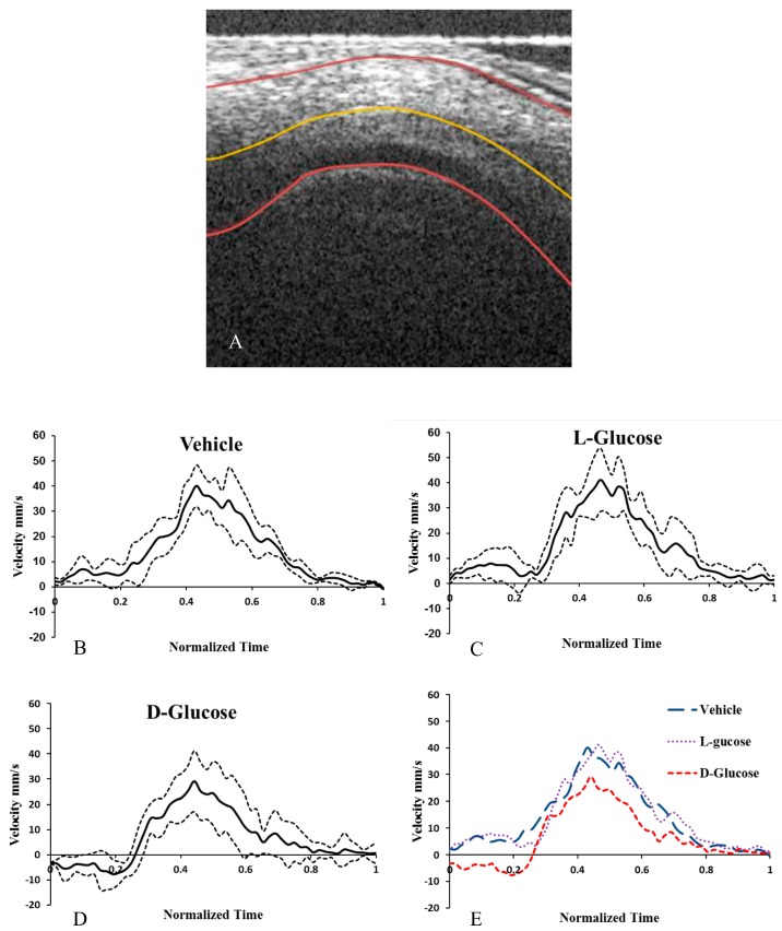
Congenital heart defects (CHDs) represent the most common form of human birth defects; approximately one-third of heart defects involve malformations of the outflow tract (OFT). Maternal diabetes increases the risk of CHD by 3-5 fold. During heart organogenesis, little is known about the effects of hyperglycemia on hemodynamics, which are critical to normal heart development. Heart development prior to septation in the chick embryo was studied under hyperglycemic conditions. Sustained hyperglycemic conditions were induced, raising the average plasma glucose concentration from 70 mg/dL to 180 mg/dL, akin to the fasting plasma glucose of a patient with diabetes. The OFTs were assessed for structural and hemodynamic alterations using optical coherence tomography (OCT), confocal microscopy, and microcomputed tomography. In hyperglycemic embryos, the endocardial cushions of the proximal OFT were asymmetric, and the OFTs curvature and torsion were significantly altered. The blood flow velocity through the OFT of hyperglycemic embryos was significantly decreased, including flow reversal in 30% of the cardiac cycle. Thus, hyperglycemia at the onset of gestation results in asymmetric proximal endocardial cushions, abnormal OFT curvature, and altered hemodynamics in the developing heart. If present in humans, these results may identify early developmental alterations that contribute to the increased risk for cardiac malformations in babies from diabetic mothers.

Keywords: congenital heart defect; development; diabetes; hemodynamics.


Conflict of interest statement

The authors declare no conflict of interest. The founding sponsors had no role in the design of the study; in the collection, analyses, or interpretation of data; in the writing of the manuscript, and in the decision to publish the results.

Figures

Figure 1
Figure 1
Hyperglycemia can lead to asymmetric endocardial outflow tract (OFT) cushions visible in the displaced lumen of cross-sectional images. Cross-sectional optical coherence tomography (OCT) images of the OFT showing a vehicle control (A), a vehicle control cross section labelled for reference (B), an osmotic control (C), a d-glucose-treated embryo with the lumen displaced towards the ventral portion of the OFT (D). Scale bar = 70 µm.
Figure 2
Figure 2
Hyperglycemia induces asymmetric endocardial OFT cushions at Hamburger–Hamilton stage 24 (HH24). Representative confocal images of a slice of the OFT cushions taken in the sagittal plane cushions labeled, and asterisks marking the two endocardial cushions from a representative vehicle control (A), an osmotic control (B), and a d-glucose-treated embryo (C). Scale bar = 100 µm, equivalent across images.
Figure 3
Figure 3
Segmented HH24 OFTs indicate endocardial OFT cushion asymmetry in response to hyperglycemia. Representative, reconstructed microcomputed tomography (Micro-CT) images of the OFT (transparent purple) with the cushions, left inferior, right superior, (purple) identified for a vehicle control (A), an osmotic control (B), and a d-glucose-treated embryo (C). (D) Average percentage difference in OFT cushion volume. Scale bar = 100 µm, equivalent across images. * p < 0.05 ANOVA.
Figure 4
Figure 4
Micro-CT analysis of HH24 hearts reveals differences in endocardial OFT properties in response to hyperglycemia. Representative, reconstructed micro-CT images of the atria (white), ventricle (dark purple), and OFT (light purple) for a vehicle control (A), an osmotic control (B), and a d-glucose-treated embryo (C). The average surface area (D) and volume (E) for the heart sections. Scale bar = 100 µm. * p < 0.05 ANOVA.
Figure 5
Figure 5
Hyperglycemic conditions can result in changes in the torsion of the OFT. 3D centerlines of a HH24 representative (A) vehicle control embryo and (B) d-glucose-treated embryo to quantify the amount of torsion of the OFT. (CE) Graphs, separated by treatment, of the average cumulative torsion, with the dashed lines representing + and −1 standard deviations of the OFT torsion plotted against the cumulative arc length. (F) The average cumulative torsion of the three treatments plotted against the average arc length.
Figure 6
Figure 6
Hyperglycemia alters the hemodynamics in chick embryonic OFT. OCT image of the longitudinal cross section of a vehicle control HH18 OFT with lumen and myocardium labelled (A). The outer walls and centerline of the OFT identified with the custom MATLAB program. The calculated centerline velocity of the vehicle control (B), osmotic control (C), and d-glucose-treated embryos (D) from the Doppler OCT images. The average velocities (E) for the treatments. Scale bar = 70 µm. * p < 0.05 ANOVA.

References

    1. Hornberger L.K. Maternal Diabetes and the Fetal Heart. Heart. 2006;92:1019–1021. doi: 10.1136/hrt.2005.083840. - DOI - PMC - PubMed
    1. Kousseff B.G. Gestational Diabetes Mellitus (Class A): A Human Teratogen? Am. J. Med. Genet. Part A. 1999;83:402–408. doi: 10.1002/(SICI)1096-8628(19990423)83:5<402::AID-AJMG12>3.0.CO;2-F. - DOI - PubMed
    1. Kousseff B.G. Diabetic Embryopathy. Curr. Opin. Pediatr. 1999;11:348–352. doi: 10.1097/00008480-199908000-00014. - DOI - PubMed
    1. Jenkins K.J., Correa A., Feinstein J.A., Botto L., Britt A.E., Daniels S.R., Elixson M., Warnes C.A., Webb C.L. Noninherited Risk Factors and Congenital Cardiovascular Defects: Current Knowledge—A Scientific Statement from the American Heart Association Council on Cardiovascular Disease in the Young. Circulation. 2007;115:2995–3014. doi: 10.1161/CIRCULATIONAHA.106.183216. - DOI - PubMed
    1. Abu-Daya A., Sater A.K., Wells D.E., Mohun T.J., Zimmerman L.B. Absence of Heartbeat in the Xenopus Tropicalis Mutation Muzak Is Caused by a Nonsense Mutation in Cardiac Myosin myh6. Dev. Biol. 2009;336:20–29. doi: 10.1016/j.ydbio.2009.09.019. - DOI - PMC - PubMed

LinkOut - more resources