. 2018 Feb 13;49(1):15.
doi: 10.1186/s13567-018-0508-z.

SeMet attenuates OTA-induced PCV2 replication promotion by inhibiting autophagy by activating the AKT/mTOR signaling pathway

Affiliations

SeMet attenuates OTA-induced PCV2 replication promotion by inhibiting autophagy by activating the AKT/mTOR signaling pathway

Gang Qian et al. Vet Res. .

Abstract

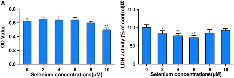
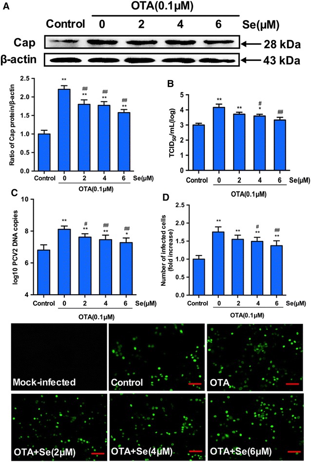
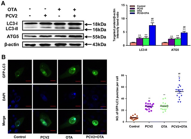
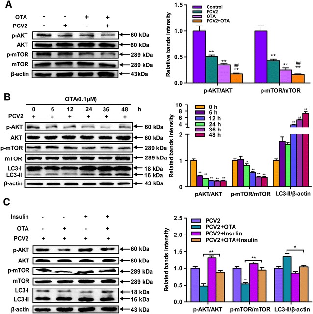
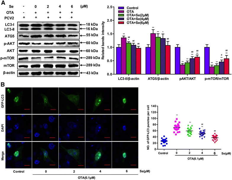
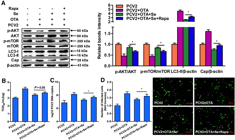
Porcine circovirus type 2 (PCV2) is recognized as the causative agent of porcine circovirus-associated diseases. PCV2 replication could be promoted by low doses of ochratoxin A (OTA) as in our previous study and selenium has been shown to attenuate PCV2 replication. However, the underlying mechanism remains unclear. The aim of the study was to investigate the effects of selenomethionine (SeMet), the major component of organic selenium, on OTA-induced PCV2 replication promotion and its potential mechanism. The present study demonstrates that OTA could promote PCV2 replication as measured by cap protein expression, viral titer, viral DNA copies and the number of infected cells. In addition, OTA could activate autophagy as indicated by up-regulated light chain 3 (LC3)-II and autophagy-related protein 5 expressions and autophagosome formation. Further, OTA could down-regulate p-AKT and p-mTOR expressions and OTA-induced autophagy was inhibited when insulin was applied. SeMet at 2, 4 and 6 μM had significant inhibiting effects against OTA-induced PCV2 replication promotion. Furthermore, SeMet could attenuate OTA-induced autophagy and up-regulate OTA-induced p-AKT and p-mTOR expression inhibition. Rapamycin, an inhibitor of AKT/mTOR, could reverse the effects of SeMet on OTA-induced autophagy and the PCV2 replication promotion. In conclusion, SeMet could block OTA-induced PCV2 replication promotion by inhibiting autophagy by activating the AKT/mTOR pathway. Therefore, SeMet supplementation could be an effective prophylactic strategy against PCV2 infections and autophagy may be a potential marker to develop novel anti-PCV2 drugs.


Figures

Figure 1
Figure 1
Effects of selenium on cell viability and LDH activity in PK-15 cells. PK-15 cells were seeded in 96-well plates at a density of 5 × 103 cells/well and cultured with selenium at various concentrations for 72 h. Cell viability (A) and LDH activity (B) were determined as described in the materials and methods. Data are presented as mean ± SE of three independent experiments. Significance compared with control (0 μM Se), *P < 0.05 and **P < 0.01.
Figure 2
Figure 2
Selenium blocks the PCV2 replication promoted by OTA. PK-15 cells were cultured for 12 h with selenium at various concentrations and then treated with PCV2 in the presence or absence of 0.1 μM OTA for another 60 h. Cells were assayed for A PCV2 viral cap protein expression by Western blotting, B PCV2 viral DNA copies by Quantitative real-time PCR, C virus titers and D the number of infected cells (Scar bar: 100 μm) by IFA as described in Materials and methods. Data are presented as mean ± SE of three independent experiments. Significance compared with control, *P < 0.05 and **P < 0.01. Within the OTA-treated groups, significance compared with cells without selenium, #P < 0.05 and ##P < 0.01.
Figure 3
Figure 3
Effects of OTA treatment on autophagy in PCV2-infected PK-15 cells. A PK-15 cells were infected with or without PCV2 for 24 h then incubated in the presence or absence of 0.1 μM OTA for 48 h. After harvest, the expressions of LC3, ATG5 and β-actin were analyzed by Western blotting as described in Materials and methods. Data are presented as mean ± SE of three independent experiments. Significance compared with control, *P < 0.05 and **P < 0.01. Significance compared with PCV2, #P < 0.05 and ##P < 0.01. B PK-15 cells were transfected with GFP-LC3 plasmid. After 12 h, PK-15 cells were infected with or without PCV2 for 24 h then incubated in the presence or absence of 0.1 μM OTA for 48 h and the fluorescence signals were visualized by confocal immunofluorescence microscopy (Scale bar: 10 μm). The average number of LC3 puncta in each cell was determined from at least 100 cells in each group. Data are presented as mean ± SE of three independent experiments. Significance compared with control, *P < 0.05 and **P < 0.01. Significance compared with PCV2, #P < 0.05 and ##P < 0.01.
Figure 4
Figure 4
OTA treatment induce autophagy by suppressing AKT/mTOR signaling pathway. A PK-15 cells were infected with PCV2 for 24 h then incubated with 0.1 μM OTA for 48 h. Cells were harvested and the expressions of p-AKT, AKT, p-mTOR, mTOR, LC3 and β-actin were analyzed by Western blotting as described in Materials and methods. Data are presented as mean ± SE of three independent experiments. Significance compared with control, *P < 0.05 and **P < 0.01. Significance compared with PCV2, #P < 0.05 and ##P < 0.01. B PK-15 cells were infected with PCV2 for 24 h then incubated with 0.1 μM OTA. Cells were harvested at the indicated time and the expressions of p-AKT, AKT, p-mTOR, mTOR, LC3 and β-actin were analyzed by Western blotting as described in Materials and methods. Data are presented as mean ± SE of three independent experiments. Significance compared with control, *P < 0.05 and **P < 0.01. C PK-15 cells were infected with PCV2 for 24 h then incubated with 0.1 μM OTA in the presence or absence of 100 μg/mL insulin for 48 h. After harvest, the expressions of p-AKT, AKT, p-mTOR, mTOR, LC3 and β-actin were analyzed by Western blotting as described in materials and methods. Data are presented as mean ± SE of three independent experiments. *P < 0.05 and **P < 0.01.
Figure 5
Figure 5
Effects of selenium supplementation on OTA-induced autophagy in PCV2-infected PK-15 cells. A PK-15 cells were cultured with or without selenium for 12 h then incubated with PCV2 and 0.1 μM OTA for another 60 h. After harvest, the expressions of LC3, ATG5, p-AKT, AKT, p-mTOR, mTOR and β-actin were analyzed by Western blotting as described in Materials and methods. Data are presented as mean ± SE of three independent experiments. Significance compared with control, *P < 0.05 and **P < 0.01. Significance compared with OTA, #P < 0.05 and ##P < 0.01. B PK-15 cells were transfected with GFP-LC3 plasmid. Then PK-15 cells were cultured with or without selenium for 12 h and incubated with PCV2 and 0.1 μM OTA for another 60 h and the fluorescence signals were visualized by confocal immunofluorescence microscopy (Scale bar: 10 μm). The average number of LC3 puncta in each cell was determined from at least 100 cells in each group. Data are presented as mean ± SE of three independent experiments. Significance compared with control, *P < 0.05 and **P < 0.01. Significance compared with PCV2, #P < 0.05 and ##P < 0.01.
Figure 6
Figure 6
Inactivation of AKT/mTOR signaling pathway abolishes the inhibitory effect of selenium on OTA-induced autophagy and PCV2 replication promotion. PK-15 cells were pretreated with rapamycin and cultured with or without selenium for 12 h before incubation with PCV2 and 0.1 μM OTA for another 60 h. After harvest, A expressions of LC3, Cap, p-AKT, AKT, p-mTOR, mTOR and β-actin were analyzed by Western blotting. B Virus titers by IFA. C PCV2 viral DNA copies by Quantitative real-time PCR. D the number of infected cells (Scar bar: 100 μm) by IFA as described in materials and methods. Data are presented as mean ± SE of three independent experiments. *P < 0.05, **P < 0.01.

Similar articles

Cited by

References

    1. Tischer I, Gelderblom H, Vettermann W, Koch MA. A very small porcine virus with circular single-stranded DNA. Nature. 1982;295:64–66. doi: 10.1038/295064a0. - DOI - PubMed
    1. Todd D, Bendinelli M, Biagini P, Hino S, Mankertz A, Mishiro S, Niel C, Okamoto H, Raidal S, Ritchie BW, Teo GC (2005) Virus taxonomy, 8th report of the International Committee for the taxonomy of viruses. pp 327–334
    1. Segalés J, Domingo M. Postweaning multisystemic wasting syndrome (PMWS) in pigs. A review. Vet Q. 2002;24:109–124. doi: 10.1080/01652176.2002.9695132. - DOI - PubMed
    1. Kim J, Chung HK, Chae C. Association of porcine circovirus 2 with porcine respiratory disease complex. Vet J. 2003;166:251–256. doi: 10.1016/S1090-0233(02)00257-5. - DOI - PubMed
    1. Jensen TK, Vigre H, Svensmark B, Bille-Hansen V. Distinction between porcine circovirus type 2 enteritis and porcine proliferative enteropathy caused by Lawsonia intracellularis. J Comp Pathol. 2006;135:176–182. doi: 10.1016/j.jcpa.2006.08.006. - DOI - PubMed

Publication types

MeSH terms

LinkOut - more resources