. 2018 Feb 13;37(1):28.
doi: 10.1186/s13046-018-0693-7.

MYL6B, a myosin light chain, promotes MDM2-mediated p53 degradation and drives HCC development

Affiliations

MYL6B, a myosin light chain, promotes MDM2-mediated p53 degradation and drives HCC development

Xingwang Xie et al. J Exp Clin Cancer Res. .

Abstract

Background: Identification of novel MDM2 or p53 binding proteins may reveal undefined oncogenes, tumor suppressors, signaling pathways and possible treatment targets.

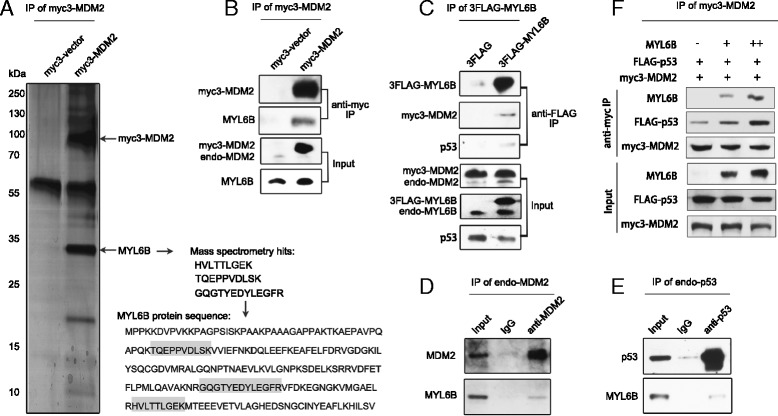
Methods: By means of immunoprecipitation and Mass Spectrometry analysis, we aimed to identify novel regulators of the MDM2-p53 pathway. We further clarified the impact of MYL6B on the p53 protein level and on the process of apoptosis. We also investigated the role of MYL6B in hepatocellular carcinoma by clone formation assay and by determining the correlation between its expression and prognosis of HCC patients.

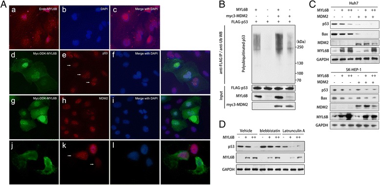
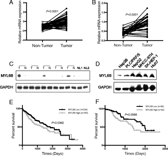
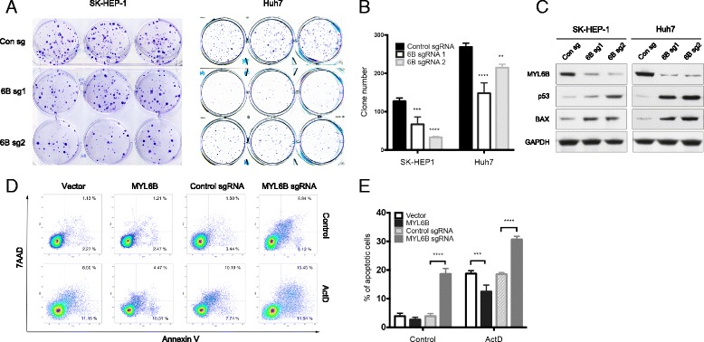
Results: We identified a novel MDM2 and p53 binding protein, MYL6B. It is a myosin light chain that could bind myosin II heavy chains to form non-muscle myosin II holoenzymes (NMII). We found that MYL6B could facilitate the binding of MDM2 to p53, which consequently promotes the ubiquitination and degradation of p53 protein. We further proved that MYL6B exerts the suppression effect on p53 as part of NMII holoenzymes because inhibiting the ATPase activity of myosin II heavy chain largely blocked this effect. We also discovered that MYL6B is overexpressed in HCC tissues and linked to the bad prognosis of HCC patients. Knocking out of MYL6B dramatically suppressed the clonogenic ability and increased the apoptosis level of HCC cell lines.

Conclusions: To summary, our results demonstrate that MYL6B is a putative tumor driver gene in HCC which could promote the degradation of p53 by enhancing its' MDM2-mediated ubiquitination.


Conflict of interest statement

Competing interest

The authors declare that they have no competing interests.

Publisher’s Note

Springer Nature remains neutral with regard to jurisdictional claims in published maps and institutional affiliations.

Figures

Fig. 1
Fig. 1
MYL6B interacts with both MDM2 and p53. (a, b) Whole cell lysate from Huh7 cell transfected with myc3-vector or myc3-MDM2 were subjected to Co-IP with anti-myc agarose and followed by NanoLC-ESI-MS/MS analysis (a) or immunoblotting analysis with antibody against MDM2 or MYL6B (B). (c) Whole cell lysate from Huh7 cell transfected with 3FLAG-vector or 3FLAG-MYL6B were subjected to Co-IP with anti-FLAG Agarose Affinity Gel and followed by immunoblotting with antibody against MYL6B, MDM2 or p53. (d) Huh7 cell lysate was subjected to Co-IP with anti-MDM2 or IgG and followed by immunoblotting with antibody against MYL6B. (e) Whole cell lysate from Huh7 cell lysate was subjected to Co-IP with anti-p53 or IgG and followed by immunoblotting with antibody against MYL6B. (f) Increasing amounts (0, 1, and 2 μg) of MYL6B plasmid were transfected into Huh7 together with myc3-MDM2 and FLAG-p53. Anti-myc was used to immunoprecipitate myc3-MDM2 and immunoblotting were performed with p53, MYL6B and MDM2 antibodies
Fig. 2
Fig. 2
MYL6B negatively regulates p53 by promoting its’ ubiquitination. (a) Huh7 was processed for immunofluorescence analysis with anti-MYL6B (a-c). Huh7 transfected with Myc-DDK-MYL6B was subjected for immunofluorescence analysis with anti-FLAG and combing with either anti-p53 (d-i) or anti-MDM2 (j-l). DAPI (4′,6-diamidino-2-phenylindole) was used to label nuclei. (b) Huh7 was cotransfected with FLAG-p53, MYL6B and myc3-MDM2. Forty-eight hours later, cells were treated with 20 μM MG132 for 4 h and harvested for immunoprecipitation assay with anti-FLAG Agarose Affinity Gel. The eluted proteins were then subjected to immunoblot analysis with ubiquitin, p53, MYL6B and MDM2 antibodies. (c) Increasing amounts (0, 1, and 2 μg) of MYL6B plasmid were transfected into Huh7 (upper panel) and SK-HEP-1 (lower panel) alone or together with MDM2 plasmid, and followed by immunoblotting with p53, Bax, MDM2, MYL6B and GAPDH antibodies. (d) Huh7 transfected with increasing amounts (0, 1, and 2 μg) of MYL6B plasmid was treated with blebbistatin (4 μM), latrunculin A (2 μM) or vehicle and then subjected to immunoblot analysis with p53, Bax, MDM2, MYL6B and GAPDH antibodies
Fig. 3
Fig. 3
MYL6B is overexpressed in HCC and linked to bad prognosis. (a, b) Total protein extraction from HCC tissues, paired non-tumor adjacent tissues and HCC cell lineswere subject to immunoblotting with MYL6B and GAPDH antibodies. (c, d) The mRNA expression level of MYL6B in HCC tissues, paired non-tumor adjacent tissues based on RNA sequencing data (RNA-seq) from the TCGA database and its’ correlation with patient prognosis. (e, f) The mRNA expression level of MYL6B in HCC tissues, paired non-tumor adjacent tissues based on real time PCR analysis of Guilin Cohort (35 patients) and its’ correlation with patient prognosis
Fig. 4
Fig. 4
MYL6B oncogene dependency in human HCC cell lines. (a, b) SK-HEP-1 and Huh7 were infected with MYL6B specific sgRNA (6B sg1 and 6B sg2) and control sgRNA. Clone formation assay were used to test the oncogene dependency of MYL6B in both cell lines, and relative clone number was used for statistic analysis. c Immunoblotting was performed after MYL6B knocking out with MYL6B, p53, BAX and GAPDH antibodies. d After MYL6B over-expressed or knocked out in Huh7, Annexin-V and 7AAD staining was used in flow cytometry analysis to measure the apoptosis level. (e) The frequency of Annein-V positive cells was indicated. *p < 0.05, ***p < 0.001, ****p < 0.0001

References

    1. Levine AJ, Oren M. The first 30 years of p53: growing ever more complex. Nat Rev Cancer. 2009;9(10):749–758. doi: 10.1038/nrc2723. - DOI - PMC - PubMed
    1. Fujimoto A, Furuta M, Totoki Y, Tsunoda T, Kato M, Shiraishi Y, Tanaka H, Taniguchi H, Kawakami Y, Ueno M, et al. Whole-genome mutational landscape and characterization of noncoding and structural mutations in liver cancer. Nat Genet. 2016;48(5):500–509. doi: 10.1038/ng.3547. - DOI - PubMed
    1. Honda R, Tanaka H, Yasuda H. Oncoprotein MDM2 is a ubiquitin ligase E3 for tumor suppressor p53. FEBS Lett. 1997;420(1):25–27. doi: 10.1016/S0014-5793(97)01480-4. - DOI - PubMed
    1. Montes de Oca Luna R, Wagner DS, Lozano G. Rescue of early embryonic lethality in mdm2-deficient mice by deletion of p53. Nature. 1995;378(6553):203–206. doi: 10.1038/378203a0. - DOI - PubMed
    1. Riley MF, Lozano G. The many faces of MDM2 binding partners. Genes & cancer. 2012;3(3–4):226–239. doi: 10.1177/1947601912455322. - DOI - PMC - PubMed

MeSH terms

Substances