. 2018 Feb 13;49(1):16.
doi: 10.1186/s13567-018-0513-2.

A novel CAV derived cell-penetrating peptide efficiently delivers exogenous molecules through caveolae-mediated endocytosis

Affiliations

A novel CAV derived cell-penetrating peptide efficiently delivers exogenous molecules through caveolae-mediated endocytosis

Gaowei Hu et al. Vet Res. .

Abstract

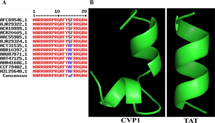
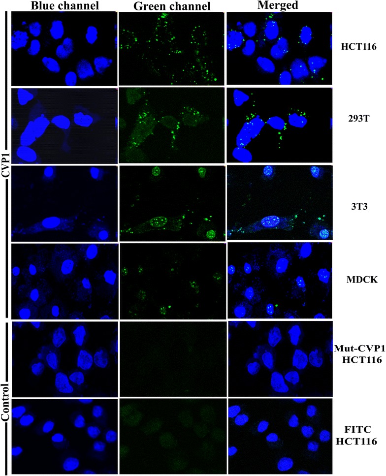
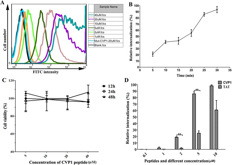
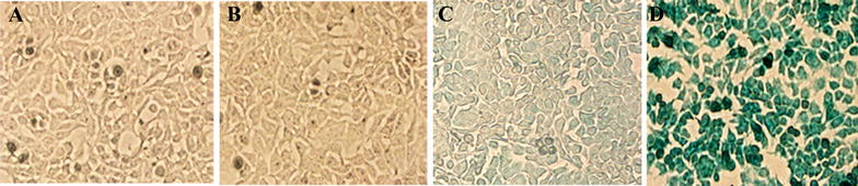
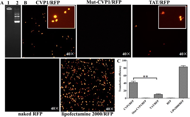
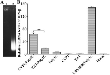
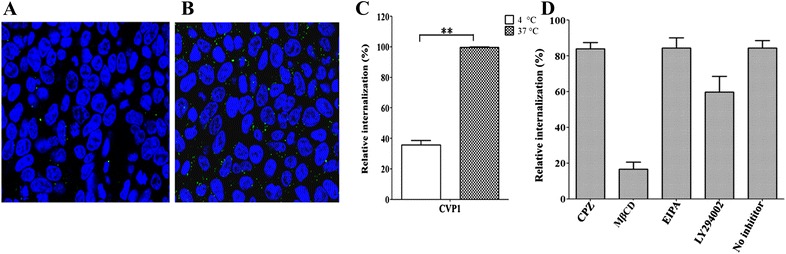
Cell-penetrating peptide (CPP) is a promising cargo for delivering bioactive molecules. In this study, the N terminus of VP1 from chicken anemia virus, designated as CVP1, was found to carry enriched arginine residues with α-helix. By confocal imaging, flow cytometry and MTT assay, we identified CVP1 as a novel, safe and efficient CPP. CVP1-FITC peptide could entry different types of cells tested with dose dependence, but without cytotoxic effects. Compared with TAT-FITC peptide, the CVP1-FITC peptide showed much higher cell-penetrating activity. Moreover, CVP1 could successfully deliver β-glycosidase, poly (I:C) and plasmid into HCT116 cells. Inhibitors and temperature sensitivity analysis further indicated that the cell-penetrating activity of CVP1 was based on ATP-dependent and caveolae-mediated endocytosis. All these data demonstrate that CVP1 has efficient cell-penetrating activity and great potential for developing a novel delivery vector.


Figures

Figure 1
Figure 1
Multi alignment of reference VP1 partial sequences and displaying the predicted structure of CVP1 and TAT by PyMOL™. A The reference strains of CAV VP1 were aligned using MultAlin server. The red letters represent the identical amino acid residues among the different reference VP1 sequences. The blue or black letters represent the different amino acid residues. B Comparison of structure between CVP and TAT.
Figure 2
Figure 2
The intracellular location of CVP1 peptide in different cell types and imaging by confocal microscopy. HCT116, 293T, 3T3, MDCK cells were treated with 5 µM of CVP1 peptide for 30 min at 37 °C. The co-incubation of Mut-CVP1 and FITC dye with HCT116 cells served as the negative control. The nuclei was stained with Hoechst 33342 and presented the blue signal. The intracellular location of FITC-labeled CVP1 was indicated by the green signal. The pictures from left to right were blue channel, green channel and overlapped images, respectively.
Figure 3
Figure 3
The character of CVP1 peptide cell-penetrating activity. A Analysis of the intake of CVP1 peptide in HCT116 cells at increasing concentration by flow cytometry. HCT116 cells were treated with different concentrations (1, 2, 5, 10, 20, 40 µM) of CVP1 or Mut-CVP1 peptide for 30 min at 37 °C. The data was shown using the FlowJo software. B HCT116 cells treated with the CVP1 peptide for the different incubation time points (5, 10, 15, 20, 25, 30 min) at a peptide concentration of 5 µM. C Percentage of viable HCT116 cells after treatment with CVP1 peptide at various concentrations (5, 10, 20, 40 µM) for 12, 24, and 48 h. D The cellular uptake efficiency of CVP1 and TAT peptide were evaluated by flow cytometry at 0.1, 1, 2, 5 and 10 µM. Single asterisk (*) and double asterisks (**) indicated P < 0.05 and P < 0.01, respectively.
Figure 4
Figure 4
The efficiency of CVP1 peptide delivering protein into HCT116 cells. A HCT116 cells treated with β-galactosidase enzyme alone. B Cells treated with X-gal staining buffer alone. C Cells treated with non-covalent complex of Mut-CVP1 peptide and β-galactosidase enzyme. D Cells treated with non-covalent complex of CVP1 peptide and β-galactosidase enzyme.
Figure 5
Figure 5
The efficiency of CVP1 peptide delivering DNA into HCT116 cells. A Detection of DNA and peptide complexes was conducted by electrophoresis and ethidium bromide staining. Lane 1: pCDNA3.1-RFP plasmid conjugated with CVP1 peptide; Lane 2: pCDNA3.1-RFP plasmid only; Fluorescence microscope images of the expression of the red fluorescence protein. The images obtained at a ×40 magnification. B Images of the CVP1/RFP complex, Mut-CVP1/RFP complex, TAT/RFP complex, pCDNA3.1-RFP only and transfection reagent lipofectamine 2000/RFP complex. C Quantification of the transfection efficiency using flow cytometry. Single asterisk (*) and double asterisks (**) indicated P < 0.05 and P < 0.01, respectively.
Figure 6
Figure 6
Assaying the poly (I:C) and CVP1 complexes by gel retardation electrophoresis and detection the relative mRNA levels of IFN-β by real-time PCR. A Testing the complex of poly (I:C) and CVP1 was conducted by electrophoresis and ethidium bromide staining. Lane 1: production of CVP1 peptide co-incubated with poly (I:C); Lane 2: poly (I:C) only; B The mRNA level of IFN-β obtained from the complexes of CVP1 peptide and TAT conjugated with poly (I:C), respectively, TAT and CVP1 peptide only, transfection reagent with poly (I:C) and blank cells. Single asterisk (*) and double asterisks (**) indicated P < 0.05 and P < 0.01, respectively.
Figure 7
Figure 7
Effect of temperature and endocytosis inhibitor on CVP1 internalization. Confocal images of HCT116 cells incubated with CVP1 (10 µM) at 4 °C (A) and 37 °C (B); C the flow cytometry data for the effect of temperature were indicated as mean ± SD. Single asterisk (*) and double asterisks (**) indicated P < 0.05 and P < 0.01, respectively. D The effect of inhibitors on endocytosis is shown as mean ± SD.

References

    1. Takeuchi T, Futaki S. Current understanding of direct translocation of arginine-rich cell-penetrating peptides and its internalization mechanisms. Chem Pharm Bull. 2016;64:1431–1437. doi: 10.1248/cpb.c16-00505. - DOI - PubMed
    1. Guo Z, Peng H, Kang J, Sun D. Cell-penetrating peptides: possible transduction mechanisms and therapeutic applications. Biomed Rep. 2016;4:528–534. doi: 10.3892/br.2016.639. - DOI - PMC - PubMed
    1. Dissanayake S, Denny WA, Gamage S, Sarojini V. Recent developments in anticancer drug delivery using cell penetrating and tumor targeting peptides. J Control Release. 2017;250:62–76. doi: 10.1016/j.jconrel.2017.02.006. - DOI - PubMed
    1. Ho A, Schwarze SR, Mermelstein SJ, Waksman G, Dowdy SF. Synthetic protein transduction domains: enhanced transduction potential in vitro and in vivo. Cancer Res. 2001;61:474–477. - PubMed
    1. Vivès E, Brodin P, Lebleu B. A truncated HIV-1 Tat protein basic domain rapidly translocates through the plasma membrane and accumulates in the cell nucleus. J Biol Chem. 1997;272:16010–16017. doi: 10.1074/jbc.272.25.16010. - DOI - PubMed

Publication types

Substances

LinkOut - more resources