

Osteoporotic Pelvic Fractures
- PMID: 29439771
- PMCID: PMC5817189
- DOI: 10.3238/arztebl.2018.0070
Osteoporotic Pelvic Fractures
Abstract
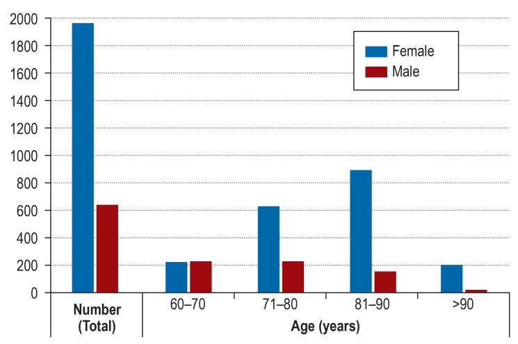
Background: The estimated incidence of osteoporotic pelvic fractures among persons over age 60 in Germany is 224 per 100 000 persons per year, and rising. A number of surgical treatment options are available, but clinical long-term data are lacking.
Methods: This review is based on pertinent publications and guidelines retrieved by a selective literature search, and on the authors' clinical experience.
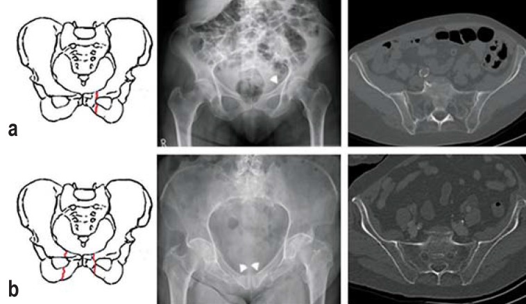
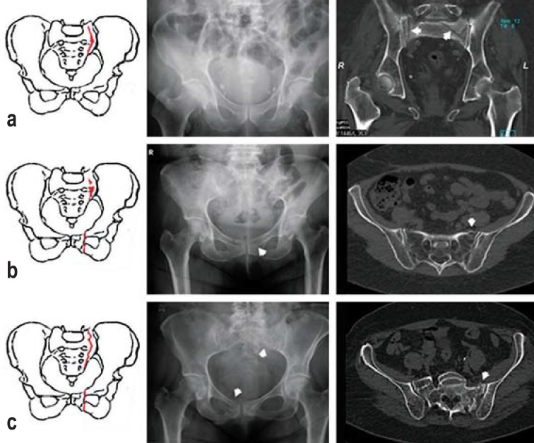
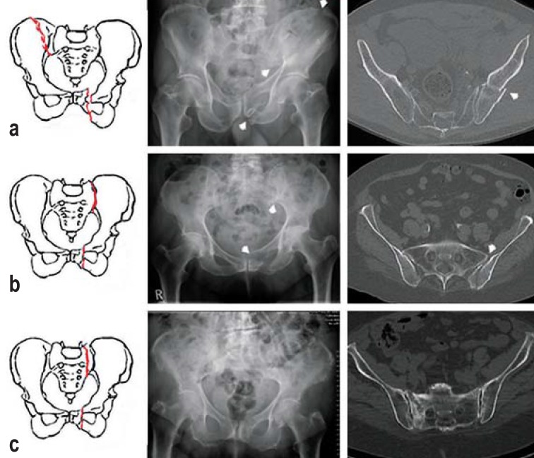
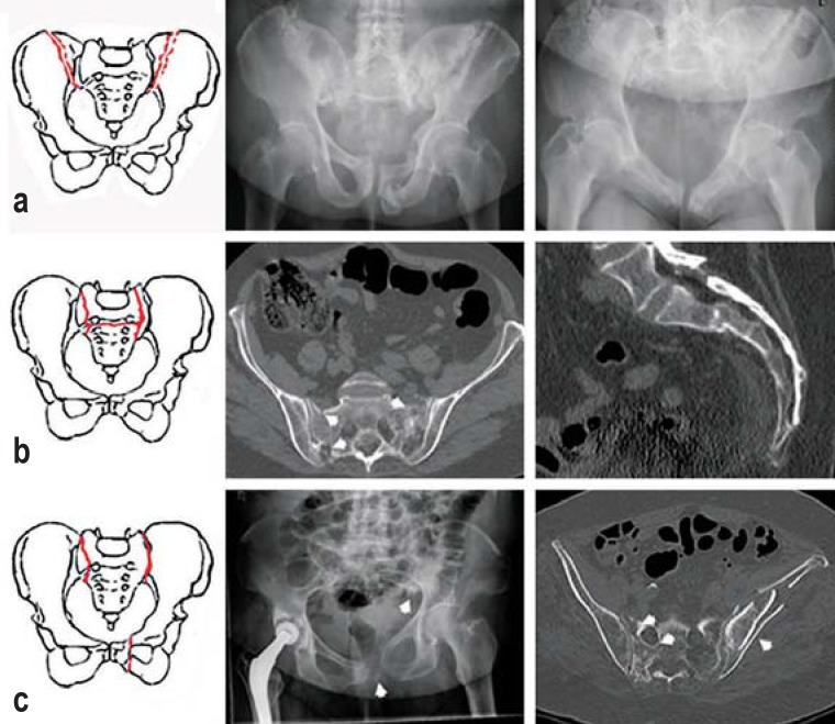
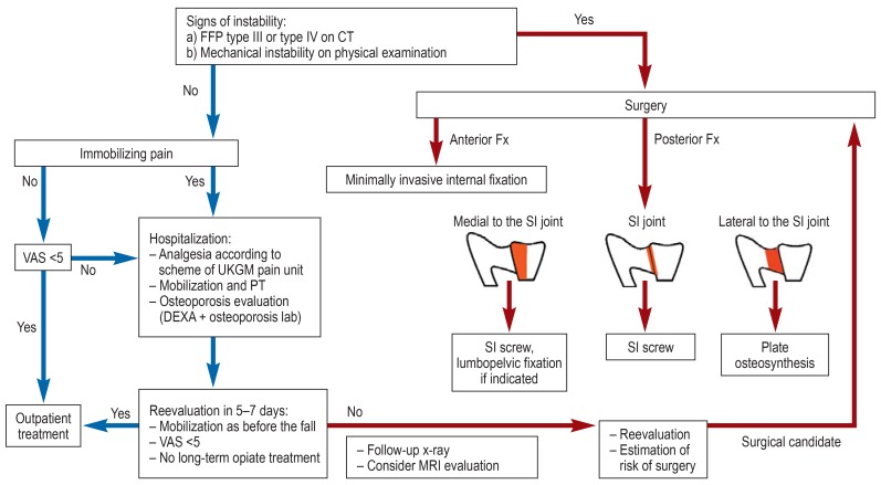
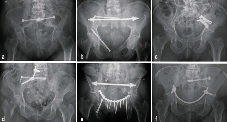
Results: Patients often report one or more relatively trivial traumatic incidents leading up to the fracture. They complain of pain in the hip, groin, or lower lumbar region, or of low back pain and sciatica. A new classification scheme entitled Fragility Fractures of the Pelvis (FFP) takes the morphology of the fracture into account and can be used as an aid to therapeutic decision-making (evidence level IV). The goal of treatment is early mobilization with adequate pain relief. Isolated anterior pelvic ring fractures (FFP I) and nondisplaced posterior pelvic ring fractures (FFP II) are usually stable and can be treated conservatively. Type III and IV injuries are unstable and should generally be treated surgically.
Conclusion: Retrospective analyses have shown that osteoporotic pelvic fractures are associated with decreased mobility and independence and with a one-year mortality ranging from 9.5% to 27%. Prospective therapeutic trials are urgently needed.
Figures
Comment in
-
CT-Guided Pelvic Osteosynthesis.Dtsch Arztebl Int. 2018 Apr 20;115(16):284. doi: 10.3238/arztebl.2018.0284a. Dtsch Arztebl Int. 2018. PMID: 29739496 Free PMC article. No abstract available.
References
-
- WHO. Guidelines for preclinical evaluation and clinical trials in osteoporosis; 1998. http://apps.who.int/iris/bitstream/10665/42088/1/9241545224_eng.pdf (last accessed on 3 Januaray 2018)
-
- Rommens PM, Wagner D, Hofmann A. Fragility fractures of the pelvis. J Bone Jt Surg Rev. 2017;5:1–13. - PubMed
-
- Krappinger D, Kammerlander C, Hak DJ, Blauth M. Low-energy osteoporotic pelvic fractures. Arch Orthop Trauma Surg. 2010;130:1167–1175. - PubMed
-
- McCabe MP, Smyth MP, Richardson DR. Current concept review: Vitamin D and stress fractures. Foot Ankle Int. 2012;33:526–533. - PubMed
Publication types
MeSH terms
LinkOut - more resources
Full Text Sources
Other Literature Sources
Medical