

The Transcriptional Regulator HlyU Positively Regulates Expression of exsA, Leading to Type III Secretion System 1 Activation in Vibrio parahaemolyticus
- PMID: 29440251
- PMCID: PMC6040195
- DOI: 10.1128/JB.00653-17
The Transcriptional Regulator HlyU Positively Regulates Expression of exsA, Leading to Type III Secretion System 1 Activation in Vibrio parahaemolyticus
Abstract
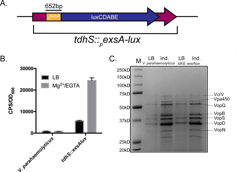
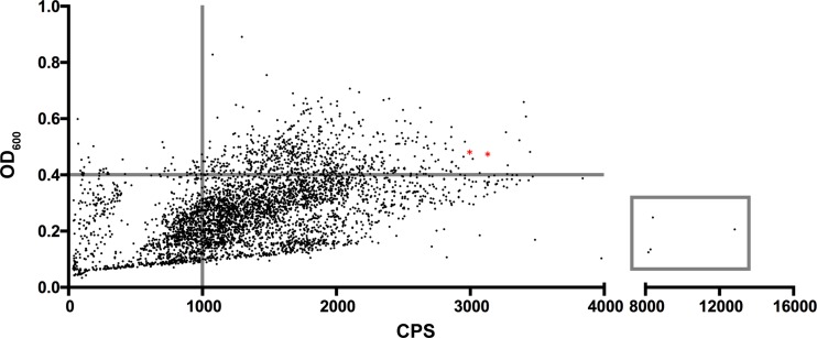
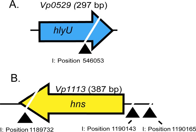
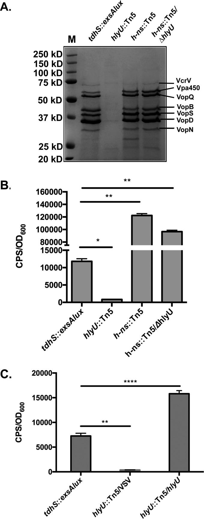
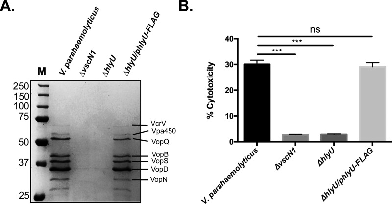
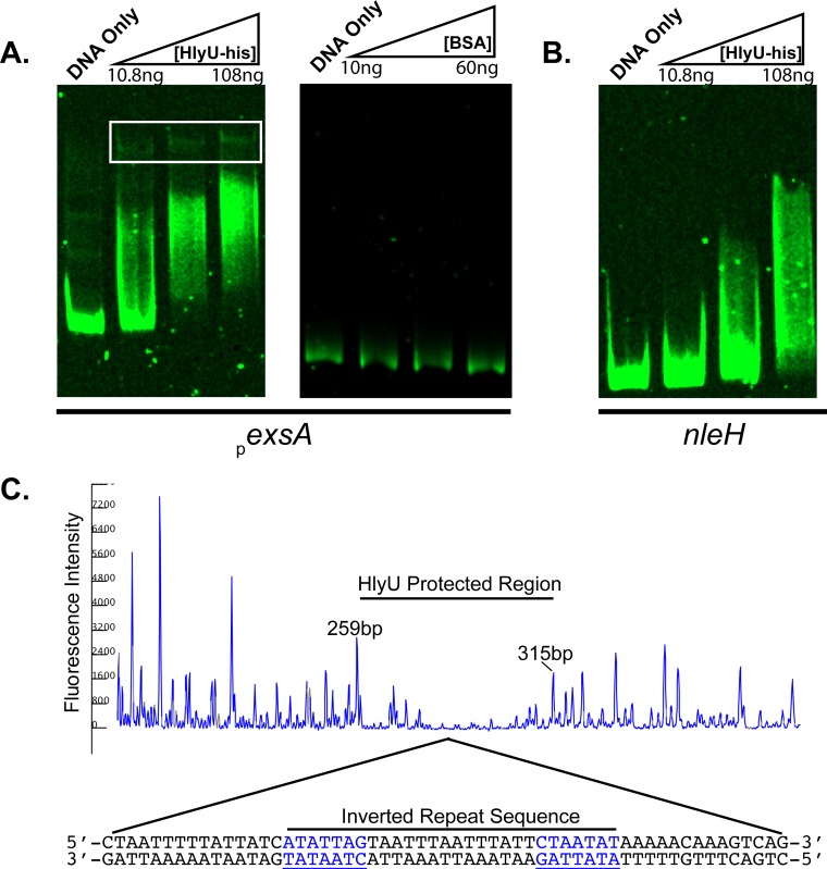
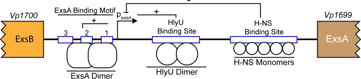
Vibrio parahaemolyticus is a marine bacterium that is globally recognized as the leading cause of seafood-borne gastroenteritis. V. parahaemolyticus uses various toxins and two type 3 secretion systems (T3SS-1 and T3SS-2) to subvert host cells during infection. We previously determined that V. parahaemolyticus T3SS-1 activity is upregulated by increasing the expression level of the master regulator ExsA under specific growth conditions. In this study, we set out to identify V. parahaemolyticus genes responsible for linking environmental and growth signals to exsA gene expression. Using transposon mutagenesis in combination with a sensitive and quantitative luminescence screen, we identify HlyU and H-NS as two antagonistic regulatory proteins controlling the expression of exsA and, hence, T3SS-1 in V. parahaemolyticus Disruption of hns leads to constitutive unregulated exsA gene expression, consistent with its known role in repressing exsA transcription. In contrast, genetic disruption of hlyU completely abrogated exsA expression and T3SS-1 activity. A V. parahaemolyticushlyU null mutant was significantly deficient for T3SS-1-mediated host cell death during in vitro infection. DNA footprinting studies with purified HlyU revealed a 56-bp protected DNA region within the exsA promoter that contains an inverted repeat sequence. Genetic evidence suggests that HlyU acts as a derepressor, likely by displacing H-NS from the exsA promoter, leading to exsA gene expression and appropriately regulated T3SS-1 activity. Overall, the data implicate HlyU as a critical positive regulator of V. parahaemolyticus T3SS-1-mediated pathogenesis.IMPORTANCE Many Vibrio species are zoonotic pathogens, infecting both animals and humans, resulting in significant morbidity and, in extreme cases, mortality. While many Vibrio species virulence genes are known, their associated regulation is often modestly understood. We set out to identify genetic factors of V. parahaemolyticus that are involved in activating exsA gene expression, a process linked to a type III secretion system involved in host cytotoxicity. We discover that V. parahaemolyticus employs a genetic regulatory switch involving H-NS and HlyU to control exsA promoter activity. While HlyU is a well-known positive regulator of Vibrio species virulence genes, this is the first report linking it to a transcriptional master regulator and type III secretion system paradigm.
Keywords: HlyU; Vibrio; enteric pathogens; gene regulation; transposons; type III secretion.
Copyright © 2018 American Society for Microbiology.
Figures
Similar articles
-
Quorum sensing employs a dual regulatory mechanism to repress T3SS gene expression.mBio. 2025 Apr 9;16(4):e0010625. doi: 10.1128/mbio.00106-25. Epub 2025 Feb 25. mBio. 2025. PMID: 39998267 Free PMC article.
-
Vfr Directly Activates exsA Transcription To Regulate Expression of the Pseudomonas aeruginosa Type III Secretion System.J Bacteriol. 2016 Apr 14;198(9):1442-50. doi: 10.1128/JB.00049-16. Print 2016 May. J Bacteriol. 2016. PMID: 26929300 Free PMC article.
-
H-NS Family Members MvaT and MvaU Regulate the Pseudomonas aeruginosa Type III Secretion System.J Bacteriol. 2019 Jun 21;201(14):e00054-19. doi: 10.1128/JB.00054-19. Print 2019 Jul 15. J Bacteriol. 2019. PMID: 30782629 Free PMC article.
-
Advances on Vibrio parahaemolyticus research in the postgenomic era.Microbiol Immunol. 2020 Mar;64(3):167-181. doi: 10.1111/1348-0421.12767. Epub 2020 Jan 21. Microbiol Immunol. 2020. PMID: 31850542 Review.
-
Fitting Pieces into the Puzzle of Pseudomonas aeruginosa Type III Secretion System Gene Expression.J Bacteriol. 2019 Jun 10;201(13):e00209-19. doi: 10.1128/JB.00209-19. Print 2019 Jul 1. J Bacteriol. 2019. PMID: 31010903 Free PMC article. Review.
Cited by
-
Spatiotemporal Regulation of Vibrio Exotoxins by HlyU and Other Transcriptional Regulators.Toxins (Basel). 2020 Aug 22;12(9):544. doi: 10.3390/toxins12090544. Toxins (Basel). 2020. PMID: 32842612 Free PMC article. Review.
-
Quorum sensing employs a dual regulatory mechanism to repress T3SS gene expression.mBio. 2025 Apr 9;16(4):e0010625. doi: 10.1128/mbio.00106-25. Epub 2025 Feb 25. mBio. 2025. PMID: 39998267 Free PMC article.
-
Transcriptomic Analysis of Vibrio parahaemolyticus Underlying the Wrinkly and Smooth Phenotypes.Microbiol Spectr. 2022 Oct 26;10(5):e0218822. doi: 10.1128/spectrum.02188-22. Epub 2022 Sep 13. Microbiol Spectr. 2022. PMID: 36098555 Free PMC article.
-
Genomic and Transcriptomic Analyses Reveal Multiple Strategies for Vibrio parahaemolyticus to Tolerate Sub-Lethal Concentrations of Three Antibiotics.Foods. 2024 May 27;13(11):1674. doi: 10.3390/foods13111674. Foods. 2024. PMID: 38890902 Free PMC article.
-
Transcriptomic Profiles of Vibrio parahaemolyticus During Biofilm Formation.Curr Microbiol. 2023 Oct 14;80(12):371. doi: 10.1007/s00284-023-03425-7. Curr Microbiol. 2023. PMID: 37838636
References
-
- Kondo H, Tinwongger S, Proespraiwong P, Mavichak R, Unajak S, Nozaki R, Hirono I. 2014. Draft genome sequences of six strains of Vibrio parahaemolyticus isolated from early mortality syndrome/acute hepatopancreatic necrosis disease shrimp in Thailand. Genome Announc 2:e00221-. doi:10.1128/genomeA.00221-14. - DOI - PMC - PubMed
Publication types
MeSH terms
Substances
LinkOut - more resources
Full Text Sources
Other Literature Sources
Molecular Biology Databases
Research Materials
Miscellaneous
