Review
. 2018 Sep 4;8(9):a031708.
doi: 10.1101/cshperspect.a031708.

Natural History, Clinical Manifestations, and Pathogenesis of Hepatitis A

Affiliations
Review

Natural History, Clinical Manifestations, and Pathogenesis of Hepatitis A

Eui-Cheol Shin et al. Cold Spring Harb Perspect Med. .

Abstract

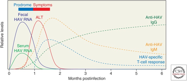
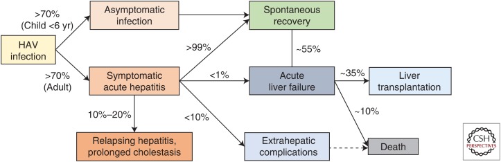
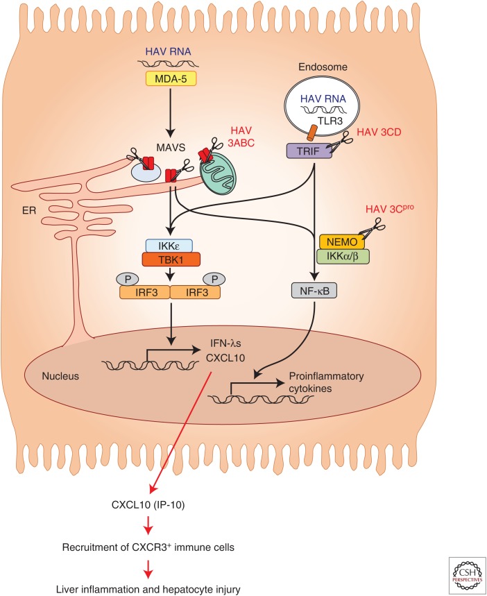
Hepatitis A virus (HAV) is transmitted by the fecal-oral route and is a major cause of acute viral hepatitis. The clinical manifestations of HAV infection range from asymptomatic infection to acute liver failure (ALF), but do not include progression to chronic hepatitis. Risk factors for severe acute hepatitis A are older age (>40 years) and preexisting liver disease. Some patients may show atypical clinical features such as relapsing hepatitis, prolonged cholestasis, or extrahepatic manifestations. Almost all hepatitis A patients spontaneously recover with supportive care. However, in the case of ALF (<1%), intensive care and urgent decision on liver transplantation are required. Liver injury during hepatitis A is not directly caused by HAV but is known to be caused by immune-mediated mechanisms. In this review, the natural history and clinical manifestations of hepatitis A are described. In addition, mechanisms of immunopathogenesis in hepatitis A are discussed.


Figures

Figure 1.
Figure 1.
A typical course of hepatitis A. After a 3- to 5-week incubation period following hepatitis A virus (HAV) infection, patients develop symptoms of hepatitis with elevation of serum alanine aminotransferase (ALT) levels. Fecal virus shedding and viremia are present and peak during the incubation period. Anti-HAV antibodies appear in serum first as immunoglobulin (Ig)M and subsequently as IgG. Virus-specific T-cell responses coincide with the elevation of serum ALT levels.
Figure 2.
Figure 2.
The clinical outcomes of hepatitis A virus (HAV) infection. Clinical manifestations of HAV infection depend on the age of patients. Most adult patients develop symptomatic hepatitis, whereas most young children do not. Common hepatitis symptoms are fever, malaise, nausea or vomiting, abdominal discomfort, and dark urine and jaundice. Reported extrahepatic complications include acute kidney injury, acalculous cholecystitis, pancreatitis, pleural or pericardial effusion, hemolysis, hemophagocytosis, pure red-cell aplasia, acute reactive arthritis, skin rash, and neurological manifestations such as mononeuritis, Guillain–Barré syndrome, and transverse myelitis.
Figure 3.
Figure 3.
Radiological findings of hepatitis A. (A) Transabdominal ultrasonography shows diffuse wall thickening of the gallbladder (arrows) measuring ∼10 mm. (B) Transverse computed tomography (CT) scan depicts low attenuating halo around the right portal vein indicating periportal tracking (arrowheads). Perihepatic lymph node enlargement is also noted (arrow).
Figure 4.
Figure 4.
Two different forms of infectious hepatitis A virus (HAV) virions. Quasi-enveloped HAV (eHAV) is detected in serum and plasma of the infected host, whereas nonenveloped, naked HAV is shed in feces. New replicated progeny virus is released from hepatocytes in the quasi-enveloped form and subsequently loses its lipid envelope following exposure to bile salts in the biliary canaliculus. The viral capsid within the quasi-enveloped eHAV virion is protected from neutralizing antibodies. Nonenveloped, naked HAV is highly stable. It is shed in feces via the intestinal tract and maintains infectivity in the environment. LSECs, Liver sinusoidal endothelial cells.
Figure 5.
Figure 5.
Mechanism of CXC-chemokine ligand (CXCL)10 expression in Hepatitis A virus (HAV)-infected hepatocytes. HAV RNA is sensed by MDA5 in the cytosol or TLR3 in the endosome. However, downstream signaling is interrupted by HAV proteins as described in the text. Nonetheless, interferon λ (IFN-λ) and CXCL10 are produced from HAV-infected cells, particularly at an early stage of HAV infection. CXCL10 is produced in HAV-infected cells in a MAVS- and IRF3-dependent but IFN-independent manner. CXCL10 can contribute to liver inflammation and hepatocyte injury by recruiting CXCR3+ immune cells to the HAV-infected liver.

Similar articles

Cited by

References

    1. Ajmera V, Xia G, Vaughan G, Forbi JC, Ganova-Raeva LM, Khudyakov Y, Opio CK, Taylor R, Restrepo R, Munoz S, et al. 2011. What factors determine the severity of hepatitis A-related acute liver failure? J Viral Hepat 18: e167–e174. - PMC - PubMed
    1. Amado Leon LA, de Almeida AJ, de Paula VS, Tourinho RS, Villela DA, Gaspar AM, Lewis-Ximenez LL, Pinto MA. 2015. Longitudinal study of hepatitis A infection by saliva sampling: The kinetics of HAV markers in saliva revealed the application of saliva tests for hepatitis A study. PLoS ONE 10: e0145454. - PMC - PubMed
    1. Armstrong GL, Bell BP. 2002. Hepatitis A virus infections in the United States: Model-based estimates and implications for childhood immunization. Pediatrics 109: 839–845. - PubMed
    1. Asher LV, Binn LN, Mensing TL, Marchwicki RH, Vassell RA, Young GD. 1995. Pathogenesis of hepatitis A in orally inoculated owl monkeys (Aotus trivirgatus). J Med Virol 47: 260–268. - PubMed
    1. Chen CM, Chen SC, Yang HY, Yang ST, Wang CM. 2016. Hospitalization and mortality due to hepatitis A in Taiwan: A 15-year nationwide cohort study. J Viral Hepat 23: 940–945. - PubMed

Publication types

Substances

LinkOut - more resources