Review
. 2018 Apr 6;293(14):5026-5034.
doi: 10.1074/jbc.TM117.001433. Epub 2018 Feb 13.

Structure, function, and mechanism of the core circadian clock in cyanobacteria

Affiliations
Review

Structure, function, and mechanism of the core circadian clock in cyanobacteria

Jeffrey A Swan et al. J Biol Chem. .

Abstract

Circadian rhythms enable cells and organisms to coordinate their physiology with the cyclic environmental changes that come as a result of Earth's light/dark cycles. Cyanobacteria make use of a post-translational oscillator to maintain circadian rhythms, and this elegant system has become an important model for circadian timekeeping mechanisms. Composed of three proteins, the KaiABC system undergoes an oscillatory biochemical cycle that provides timing cues to achieve a 24-h molecular clock. Together with the input/output proteins SasA, CikA, and RpaA, these six gene products account for the timekeeping, entrainment, and output signaling functions in cyanobacterial circadian rhythms. This Minireview summarizes the current structural, functional and mechanistic insights into the cyanobacterial circadian clock.

Keywords: ATPases associated with diverse cellular activities (AAA); bacterial protein kinase; bacterial signal transduction; circadian rhythm; crystallography; nuclear magnetic resonance (NMR).


Conflict of interest statement

The authors declare that they have no conflicts of interest with the contents of this article

Figures
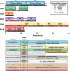
Figure 1.
Figure 1.
Domain structure, conservation, and function of core clock proteins. Domain structures of the six core clock proteins from S. elongatus are depicted as colored bars using boundaries and annotated functions from the Interpro Database (90). Sequences were aligned with homologues from the thermophilic variant T. elongatus using ClustalΩ (91). Identity scores at each position in the alignment are indicated below the domain maps as color-coded bars. Observed functions for each of the core clock protein domains are summarized below.
Figure 2.
Figure 2.
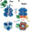
Day and night states of the cyanobacterial clock. High-resolution models of the day and nighttime states of the core oscillator are compared. Coloring scheme is the same as in Fig. 1. A, day complex. The CI and CII hexamers form stacked doughnuts (alternating subunits shown in light and dark blue for contrast, PDB code 3K0C). One subunit is shown in cartoon mode to highlight the nucleotide-binding sites (red) at the subunit interfaces. Phosphorylation of KaiC residues Thr-426 and Ser-432 takes place near the CII nucleotide-binding sites. The KaiA dimer (light and dark green for contrast) binds to C-terminal A-loops of KaiC (white, shown in complex with KaiA PDB code 5C5E). The dashed arrow represents the point of connection between the KaiA–CII loop structure and the CII A-loop extensions shown on the right. B, night complex. The intermediate-resolution cryo-EM model (PDB code 5N8Y) is combined with higher resolution models from studies on independent subcomplexes. The S431E phosphomimetic of the pS/T state of KaiC binds six molecules of KaiB (PDB code 5JWQ). This results in the recruitment and sequestration of KaiA near the KaiB–CI interface (PDB code 5JWR). Output signaling occurs through interactions between KaiB and the C-terminal PsR domain of CikA (PDB code 5JYV), as well as through interactions between KaiC and SasA at dusk, although no high-resolution structure exists for the latter.
Figure 3.
Figure 3.
Conformational changes in the core oscillator. A, CI ATP hydrolysis. High-resolution crystal structures of the CI domain of KaiC in the ATP-bound (PDB code 4LTA) and ADP-bound (PDB code 4LT9, chain C) states are overlaid. Three major structural differences are noted upon ATP hydrolysis: 1) helix bearing Phe-199 is flipped into an alternative conformation; 2) α6-α7/α8 helices are repositioned away from the nucleotide-binding site; and 3) cis peptide bond between Asp-145 and Ser-146 in the ATP-bound state is found in the trans isomer in the ADP-bound state. B, KaiA inactivation. The daytime and nighttime states of KaiA are compared. During the day, the A-loops of KaiC (blue) are bound at the dimer interface, and the interdomain linker (pink) crosses over the complex. In the Night Complex, the linker shifts to occupy the A-loop binding site. C, KaiB fold-switching. KaiB undergoes a major structural reorganization between its free and KaiC-bound forms, involving changes in both secondary and tertiary structure for the C-terminal half of the protein. Both structures are represented with rainbow coloring, starting with dark blue at the N terminus. Note that the first half of the two proteins is identical (dark blue through light green), whereas the C-terminal halves are completely different. This interconversion is thought to happen spontaneously resulting in a conformational selection mechanism for formation of the KaiBC complex.
Figure 4.
Figure 4.
Integrated timekeeping, entrainment, and output signaling functions of the cyanobacterial clock. Timekeeping, entrainment, and output signaling functions are highlighted within the oscillatory cycle of the cyanobacterial clock. Powered by ATPase activity on its CI domain, KaiC cycles through a series of phosphorylation states that is interdependent on its quaternary structure. KaiA is bound to the CII domain of KaiC during the day, stimulating phosphorylation. This process is sensitive to the ATP/ADP ratio, which peaks at midday, providing an entrainment cue. At dusk, levels of oxidized quinones rise in the cell, and the clock is entrained by this as well. Around this time, KaiC reaches the pS/pT state, and SasA binds to the CI domain to activate RpaA. CI-bound SasA is eventually competed away by KaiB. Binding of KaiB is slowed by its intrinsically unfavorable equilibrium that sequesters it in inactive states. Accumulation of KaiB in its KaiC-bound form recruits and inactivates KaiA, allowing CII dephosphorylation. The input/output protein CikA also interacts with the fold-switched form of KaiB, causing CikA to dephosphorylate RpaA, inactivating it.

References

    1. Kuhlman S. J., Craig L. M., and Duffy J. F. (2017) Introduction to chronobiology. Cold Spring Harb. Perspect. Biol. 2017, a033613 10.1101/cshperspect.a033613 - DOI - PMC - PubMed
    1. Hurley J. M., Loros J. J., and Dunlap J. C. (2016) Circadian oscillators: around the transcription-translation feedback loop and on to output. Trends Biochem. Sci. 41, 834–846 10.1016/j.tibs.2016.07.009 - DOI - PMC - PubMed
    1. Nakajima M., Imai K., Ito H., Nishiwaki T., Murayama Y., Iwasaki H., Oyama T., and Kondo T. (2005) Reconstitution of circadian oscillation of cyanobacterial KaiC phosphorylation in vitro. Science 308, 414–415 10.1126/science.1108451 - DOI - PubMed
    1. Johnson C. H., Zhao C., Xu Y., and Mori T. (2017) Timing the day: what makes bacterial clocks tick? Nat. Rev. Microbiol. 15, 232–242 10.1038/nrmicro.2016.196 - DOI - PMC - PubMed
    1. Mitsui A., Kumazawa S., Takahashi A., Ikemoto H., Cao S., and Arai T. (1986) Strategy by which nitrogen-fixing unicellular cyanobacteria grow photoautotrophically. Nature 323, 720–722 10.1038/323720a0 - DOI

Publication types

MeSH terms

Substances