. 2018 Mar 20;115(12):2988-2993.
doi: 10.1073/pnas.1722086115. Epub 2018 Feb 12.

Assembly of the membrane domain of ATP synthase in human mitochondria

Affiliations

Assembly of the membrane domain of ATP synthase in human mitochondria

Jiuya He et al. Proc Natl Acad Sci U S A. .

Abstract

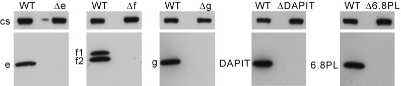
The ATP synthase in human mitochondria is a membrane-bound assembly of 29 proteins of 18 kinds. All but two membrane components are encoded in nuclear genes, synthesized on cytoplasmic ribosomes, and imported into the matrix of the organelle, where they are assembled into the complex with ATP6 and ATP8, the products of overlapping genes in mitochondrial DNA. Disruption of individual human genes for the nuclear-encoded subunits in the membrane portion of the enzyme leads to the formation of intermediate vestigial ATPase complexes that provide a description of the pathway of assembly of the membrane domain. The key intermediate complex consists of the F1-c8 complex inhibited by the ATPase inhibitor protein IF1 and attached to the peripheral stalk, with subunits e, f, and g associated with the membrane domain of the peripheral stalk. This intermediate provides the template for insertion of ATP6 and ATP8, which are synthesized on mitochondrial ribosomes. Their association with the complex is stabilized by addition of the 6.8 proteolipid, and the complex is coupled to ATP synthesis at this point. A structure of the dimeric yeast Fo membrane domain is consistent with this model of assembly. The human 6.8 proteolipid (yeast j subunit) locks ATP6 and ATP8 into the membrane assembly, and the monomeric complexes then dimerize via interactions between ATP6 subunits and between 6.8 proteolipids (j subunits). The dimers are linked together back-to-face by DAPIT (diabetes-associated protein in insulin-sensitive tissue; yeast subunit k), forming long oligomers along the edges of the cristae.

Keywords: ATP synthase; assembly; human mitochondria; membrane subunits.


Conflict of interest statement

The authors declare no conflict of interest.

Figures

Fig. 1.
Fig. 1.
Organization of subunits in monomeric ATP synthase in mammalian mitochondria. Black horizontal lines represent the inner membrane. IMS, intermembrane space. The peripheral stalk (subunits OSCP, F6, b, and d) is on the right. Subunit b has two N-terminal transmembrane α-helices interacting directly or indirectly with subunits e, f, g, DAPIT, and 6.8PL, which have no known roles in synthesis or hydrolysis of ATP. Each has a single predicted transmembrane α-helix (SI Appendix, Fig. S1). The membrane domain of subunit b is associated with mitochondrially encoded subunits ATP6 and ATP8. The N-terminal region of ATP8 has a single transmembrane α-helix, and its C-terminal region extends into the peripheral stalk. ATP8 and subunit b keep ATP6 in contact with the c8 ring. The pmf drives the rotation of this ring and the attached central stalk (subunits γ, δ, and ε) by the translocation of protons through the interface between the c8 ring and ATP6. The rotation of the central stalk carries energy into the three catalytic sites in the F1 domain (subunit composition α3β3γδε). An α subunit has been removed to expose the central γ subunit.
Fig. 2.
Fig. 2.
Absence of individual supernumerary subunits of ATP synthase from clonal HAP1 cells. Mitoplasts were prepared from HAP1-WT cells and HAP1-Δe, -Δf, -Δg, -ΔDAPIT, and -Δ6.8PL clonal cells. In the HAP1-Δf cells, f1 and f2 denote isoforms of subunit f. The mitoplasts were extracted with n-Dodecyl-β-D-maltoside. The extracts were fractionated by SDS/PAGE and Western blotted with antibodies against the corresponding subunits. Citrate synthase (cs) served as a loading control.
Fig. 3.
Fig. 3.
Subunit compositions of vestigial ATP synthase complexes produced by removal of individual supernumerary membrane subunits. The complexes were immunopurified from mitoplasts derived from HAP1-WT, -Δe, -Δf, -Δg, -ΔDAPIT, and -Δ6.8PL cells, fractionated by SDS/PAGE, and stained with Coomassie blue. The positions of subunits are indicated.
Fig. 4.
Fig. 4.
Effects of removal of individual supernumerary membrane subunits on human ATP synthase. Shown are relative abundances of subunits and forms M1, M2, and M3 of IF1 in immunopurified ATP synthase and vestigial complexes, from clonal cells. (A) HAP1-Δe. (B) HAP1-Δf. (C) HAP1-Δg. (D) HAP1-ΔDAPIT. (E) HAP1-Δ6.8PL. The histograms, derived from SI Appendix, Fig. S5 and Datasets S1–S10, are the median values of both relative abundance ratios determined for proteins found in complementary SILAC experiments. Error bars show the range of the two values. IF1-M1, -M2, and -M3 are different mature forms of IF1, and f-1 and f-2 are isoforms of subunit f (Swiss-Prot P56134). Similar experiments conducted on mitoplasts are presented in SI Appendix, Fig. S6.
Fig. 5.
Fig. 5.
Oligomeric states of ATP synthase and vestigial forms in HAP1 cells. ATP synthase and vestigial complexes were extracted at various digitonin/protein (g/g) ratios indicated above the lanes, from mitoplasts of HAP1-WT, -Δe, -Δf, -Δg, -ΔDAPIT, and -Δ6.8PL cells. Extracts were fractionated by BN-PAGE, and complexes were revealed by Western blotting with antibodies against various subunits of ATP synthase: β subunit (A), b subunit (B), γ subunit (C), and d subunit (D). Citrate synthase (cs) served as a loading control. The positions of the complexes are indicated on the left; d, dimers; m, monomers; o, oligomers; s1, F1-c8 subcomplexes; s2, subcomplexes containing subunits b, e, and g.
Fig. 6.
Fig. 6.
Pathway of assembly of the membrane domain of human ATP synthase. (AD, F, and G) Intermediate vestigial complexes and subunit compositions characterized from mitochondria of clonal cells: HAP1-Δb or -ΔOSCP (A), HAP1-Δe or -Δg (B), HAP1-Δf (C), HAP1-Δc (D), HAP1-Δ6.8PL (F), and HAP1-ΔDAPIT (G). (E) Vestigial complex from the mitochondria of ρ0 cells. The horizontal black lines denote the boundaries of the inner mitochondrial membrane. (H) The complete monomeric ATP synthase.
Fig. 7.
Fig. 7.
Structure of the dimeric Fo domain of the ATP synthase from S. cerevisiae (17). The c10 ring is in gray, and other subunits are colored and identified on the structures. In A and B, the two associated monomers are viewed from the matrix and from the intermembrane space, respectively. The interface between monomers is formed mainly by the two ATP6 (or a) subunits and by the two j subunits. Here it is proposed that subunit j and the human 6.8PL (labeled 6.8) are orthologs. It is also proposed that subunit k is the ortholog of human DAPIT, and that these subunits aid the formation of oligomeric structures consisting of rows of dimers along the edges of the mitochondrial cristae.

Comment in

  • Assembling the mitochondrial ATP synthase.
    Song J, Pfanner N, Becker T. Song J, et al. Proc Natl Acad Sci U S A. 2018 Mar 20;115(12):2850-2852. doi: 10.1073/pnas.1801697115. Epub 2018 Mar 7. Proc Natl Acad Sci U S A. 2018. PMID: 29514954 Free PMC article. No abstract available.

References

    1. Mitchell P. Coupling of phosphorylation to electron and hydrogen transfer by a chemi-osmotic type of mechanism. Nature. 1961;191:144–148. - PubMed
    1. Walker JE. The ATP synthase: The understood, the uncertain and the unknown. Biochem Soc Trans. 2013;41:1–16. - PubMed
    1. Walker JE. Structure, mechanism and regulation of ATP synthases. In: Wikström M, editor. Mechanisms of Primary Energy Transduction in Biology. Royal Society of Chemistry; London: 2017. pp. 338–373.
    1. Fearnley IM, Walker JE. Two overlapping genes in bovine mitochondrial DNA encode membrane components of ATP synthase. EMBO J. 1986;5:2003–2008. - PMC - PubMed
    1. Rak M, Gokova S, Tzagoloff A. Modular assembly of yeast mitochondrial ATP synthase. EMBO J. 2011;30:920–930. - PMC - PubMed

Publication types

Substances